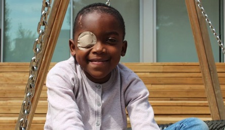
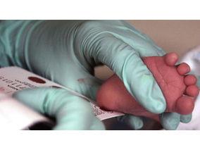
When Edwidge, a 9-year-old girl from Rwanda, arrived at SJD Barcelona Children’s Hospital for treatment, the lesion that had been growing in her right eye for years was over 5 centimetres in diameter. It was an aneurysmal bone cyst, a benign vascularised lesion that had grown excessively in her home country, as it had not previously been treated. Indeed, if this vascularisation, an excess of blood vessels surrounding the lesion, had burst, the risk of bleeding with severe complications could have been life-threatening.
Furthermore, the growth of the tumour had damaged the optic nerve, to the point that the girl had lost the sight of the right eye and had caused a deformity in it. The main objective was to remove the enormous intracranial lesion in the little girl’s orbital region and repair, as far as possible, the damage caused by the growth of the lesion. The severity of the problem, already in a very advanced state, required rapid action to prevent any complications.
This case is one of the 12 that the Probitas Foundation funds every year within the Cuida’m international cooperation programme. The surgery was studied and planned in the 3D Unit of the SJD Barcelona Children’s Hospital. In the initial phase, the lesion was analysed and, due to its vascularisation, it was considered desirable for the interventional radiology team, led by Napoleón Macías, to perform two artery embolisations before the operation. The aim was to “dry up” the tumour to stem the blood flow and prevent bleeding during resection.
Both the cutting guidelines to remove the lesion and the prosthesis to replace the bone damaged by the tumour were designed in parallel, using 3D technology. The company Avinent, also collaborated in preparing the cutting guidelines and the final prosthesis, made with PEEK (polyether ether ketone) a biocompatible material that is widely used for manufacturing prosthetics, due to its mechanical and thermal properties.
During the surgery, performed by the maxillofacial surgeon, Josep Rubio, and the neurosurgeon, Antonio Guillén, the intracranial tumour in the ocular area was cut away and the diseased bone, which could easily fracture, was removed. The custom-made prosthesis was also implanted. Over time, this will become anchored to the bone and will require no further intervention, a key element in this case, since, due to the patient’s origin, follow-up would be problematic.
Before the surgery, the stigma the girl suffered in her home country was considerable, due to the physical aspect of the lesion and the beliefs of the community in which she lives. The little girl had limitations on playing because of the risk of being hit, and doctors had warned of the risk to the girl’s life, without being able to offer her a solution in local hospitals.
Just a few months after the operation, which took place in April 2018, the impact of it on the girl’s life is clear to see. Edwidge has been able to resume normal activities, return to school and interact easily with the children and other members of her community.

AtCDF3 gene induced greater production of sugars a...
Un estudio con datos de los últimos 35 años, ind...
Un equipo de investigadores de la Universidad Juli...

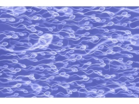
En nuestro post hablamos sobre este interesante tipo de célula del...
Palobiofarma S.L. is pleased to announce the “last patient last visi...