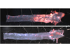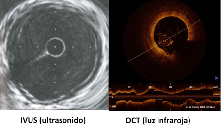
Optical Coherence Tomography (OCT) is a tool that emits infrared light. Cardiologists have been using it for about ten years to observe the inside of the coronary arteries in detail. In addition, it allows a longitudinal reconstruction of the artery. OCT has an image resolution that is 10 times higher than ultrasound, or conventional angiography, currently the most widely used ultrasound imaging technique. It allows the interior of the artery to be visualised, but also shows wall details.
The main function of OCT is to provide information on the composition of the coronary plaques. It is also an easy way to identify any plaque or coronary artery ruptures, or clots. Coronary plaques are deposits of cholesterol, calcium, and other substances. When these substances build up inside the arteries, they can block them and make them rigid. Additionally, if they break and get into the circulation, they may cause a heart attack. Plaque rupture is only visible via OCT, since the fragments are smaller than the resolution offered by an ultrasound. For these reasons, OCT has filled a gap in the diagnosis of atheromatous plaques where ultrasound was less effective.
Defining what type of plaque is in the artery is also important to decide when to implant a vascular prosthesis and to check whether this has been placed correctly. This prosthesis, called a Stent, is a small metal mesh that expands inside the artery to prevent it being blocked. Depending on whether the plaque consists of calcium or cholesterol, it will require a different treatment prior to the placing of the Stent. Implanting a Stent with the help of OCT has been shown to improve the patient’s long-term prognosis
The Institut Clinic CardioVascular (ICCV) at Hospital Clínic is a benchmark in this field, in addition to leading several studies on the use of the OCT technique. One of the most important looks at patient evolution after a Stent implant. In the lab of this unit, international students, particularly from Japan, come to learn how to use the OCT technique and how to analyse the images obtained. This makes the ICCV a benchmark in Europe for its contribution to the advancement of knowledge of atheromatous plaques using OCT, as well as its accuracy in diagnosis and treatment.

AtCDF3 gene induced greater production of sugars a...
Un estudio con datos de los últimos 35 años, ind...

Un equipo de investigadores de la Universidad Juli...

En nuestro post hablamos sobre este interesante tipo de célula del...

Theriva™ Biologics has announced the presentation of preclinical da...