Limb-girdle muscular dystrophy type 3 (LGMD D3) is a rare disease causing progressive muscle weakness, caused by point mutations in the hnRNPDL-2 protein. A member of the RNA-associated ribonucleoprotein (RNP) family, it is a little-known protein with the ability to assemble to form functional amyloid structures. Amyloids are formed by the joining of thousands of pieces of the same protein to form very stable and structured fibres (protein aggregates). Their formation is often associated with diseases such as Parkinson's and Alzheimer's, but they are also used by different organisms for functional purposes, although the number of functional amyloids described in humans is still small.
Researchers at the Universitat Autònoma de Barcelona (UAB) have determined the structure of amyloid fibres formed by the protein hnRNPDL-2 in a study published in Nature Communications. Their architecture and activity suggest that they are stable, non-toxic amyloid fibres that bind nucleic acids in their aggregated state. The results indicate that LGMD D3 could be a protein loss-of-function disease: the inability to form the amyloid structures described in the study would cause the pathology.
"Our study challenges the hypothesis that the aggregation of this protein is the cause of the disease and proposes that it is the inability to form a fibrillar structure that has been selected by evolution to bind nucleic acids that causes the pathology", says Salvador Ventura, professor of Biochemistry and Molecular Biology and researcher at the Institute of Biotechnology and Biomedicine (IBB-UAB), who led the research together with the first author of the paper, Javier Garcia-Pardo, a Juan de la Cierva-Incorporación researcher at IBB-UAB.
Researchers have determined the structure of the amyloid fibres of the hnRNPDL-2 protein using high-resolution cryo-electron microscopy (cryo-EM). This is the first structure of a human functional amyloid formed by the complete protein to be solved using this technique - previously, only structures formed by fragments of these proteins had been solved. It is also the first amyloid structure determined at high-resolution by a Spanish research team.
The structure of the protein differs from that of other pathological amyloid proteins in that it has a highly hydrophilic nucleus, which includes the amino acid associated with LGMD D3. In this case, unlike in other diseases, amyloid formation is not toxic, but necessary for the function of the protein.
The results change the concept of the origin of the disease and how it should be treated, say the researchers. "Previously, we thought that, as in many neurodegenerative diseases, LGMD D3 originated because mutations in patients caused the initially soluble protein to form aggregates and, therefore, the search for anti-aggregant molecules could be a potential therapy. Now we know that this would be a mistake, since it is the incorrect formation of the fibre that seems to trigger the disease; therefore, molecules that stabilise this structure or facilitate its formation would be the most appropriate," says Salvador Ventura.
Understanding the molecular structures of amyloids
Certain human amyloids can undergo both functional and pathological aggregation, and it is therefore necessary to understand their molecular structures in order to determine their distinctive qualities and functions. For example, RNPs similar to the one studied in this research, such as hnRNPA1 or FUS, are able to form functional amyloid fibres in response to cellular stress, but they can also harbour mutations responsible for disease. These proteins are characterised by a modular architecture, including one or more nucleic acid binding domains, together with disordered regions that are responsible for the formation of their assembly into functional or pathological amyloid structures.
"In recent years, the structures of various amyloid fibres formed by fragments of RNPs have been solved. However, these assemblies may not necessarily coincide with those adopted in the context of complete proteins, as is the case of the structure obtained for hnRNPDL-2 solved in our group," explains Salvador Ventura. "In fact, our structure differs significantly from previous ones and questions some of the assumptions that were considered valid regarding the regulation of these proteins in cells," he points out.
Special techniques for resolving functional amyloids
To resolve the structure of hnRNPDL-2 in its assembled state, the research team used the cryo-EM technique, applying special techniques to resolve amyloid structures.
In the past two years, a significant number of amyloid fibre structures have been solved with this technique, but these mainly correspond to pathological amyloids involved in systemic and neurodegenerative diseases.
"Our discovery highlights the power of cryo-EM to study the function of RNPs and the reasons for their link to disease. These proteins have been little studied until now, but they are associated with diseases such as Alzheimer's, muscular dystrophies, cancer, and neurodevelopmental and neuropsychiatric disorders. Thus, our objective now is to take advantage of the experience acquired with this technique to determine the fibrillary states of other functional amyloids and study the effect of mutations, in order to better understand their implications in health and disease," says Salvador Ventura.
The development of this new technology at the UAB will allow researchers to exploit the recently installed cryo-EM platform at the Alba synchrotron, of which the University is a partner. Solving this type of structure requires a great deal of computational power. The IBB's Protein Folding and Conformational Diseases research group led by Salvador Ventura has just acquired a high-powered computer to carry out these calculations.
The study was led by Salvador Ventura, with the participation of Javier García-Pardo, Andrea Bartolomé Nafría and Marcos Gil García from the IBB research group. The structure was solved in collaboration with Stefano Ricagno and Martino Bolognesi’s research team (Università degli Studi di Milà Statale and Unitech Nolimits Center).
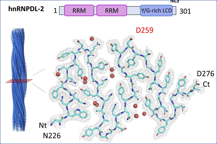
Imagen: Structure of hnRNPDL-2 amyloid fibres obtained by cryoEM at 2.5 A resolution. The upper part shows the organisation of the protein with two nucleic acid binding domains in pink and a low-complexity domain responsible for its assembly. The lower part shows the cryoEM map obtained and the structure of an amyloid fibre layer and its mutations, to better understand its implication in health and disease.
Article: Garcia-Pardo, J., Bartolomé-Nafría, A., Chaves-Sanjuan, A. et al. Cryo-EM structure of hnRNPDL-2 fibrils, a functional amyloid associated with limb-girdle muscular dystrophy D3. Nat Commun 14, 239 (2023). https://doi.org/10.1038/s41467-023-35854-0

The research team observed changes in head circumf...

AtCDF3 gene induced greater production of sugars a...
Un estudio con datos de los últimos 35 años, ind...

En nuestro post hablamos sobre este interesante tipo de célula del...
Investigadores del Cima Universidad de Navarra constatan que la combin...