A recent work of the UAB, in collaboration with the ALBA Synchrotron, has studied the side effects of typical tooth whitening treatments, based on oxidation, compared to a new treatment developed by the authors through reduction. Results showed the whitening effect of the novel treatment to be highly improved in terms of application time needed, efficiency and safety, which makes it a promising candidate to develop novel whitening treatments. Experiments at the MIRAS beamline of ALBA helped to determine the chemical mineral modifications in the dental enamel.
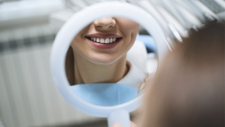
Tooth whitening is a common aesthetic treatment around the world. To obtain better results, higher concentrations of oxidizing agents and longer application times are needed, but this may increase side effects like hypersensitivity and pulp damage, tooth demineralization and gingival irritation. Besides, the need to apply these products for hours is not very comfortable for the user.
Typical tooth whitening treatments are based on the oxidizing power of hydrogen peroxide, which breaks the double bonds of the staining molecules on the teeth’s surface making them unable to absorb light. This way the molecule becomes transparent, thus obtaining a bright, clean and white smile.
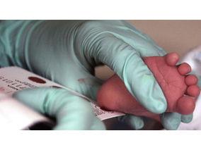
In a recent work of the Research Group of Separation Techniques in Chemistry (GTS) from the UAB in collaboration with the ALBA Synchrotron, researchers have used bovine incisors as in vitro model to study the side effects of whitening treatments. They compared typical whitening treatments (based on oxidation with carbamide peroxide) to a new treatment developed and patented by the authors through reduction via metabisulfite, which also makes the staining molecules colorless. However, metabisulfite presents a faster whitening effect, which permits the use of lower concentrations and shorter application times. Results showed how the whitening effect of the novel treatment is highly improved in terms of application time needed, with the consequent reduction of side effects. This makes it a promising candidate to develop novel whitening treatments.
“The whitening effect increased when metabisulfite is encapsulated within liposomes (vesicles made out of lipids), since it favors the diffusion of the reducing agent into the enamel, obtaining significant whitening results in 3 minutes.” explains Manuel Valiente, professor of the UAB’s Department of Chemistry and director of GTS.
In addition, they found that using oxidizing or reducing treatments promotes different mineral changes.
This was known thanks to the chemical imaging performed at the MIRAS beamline of ALBA. It is the first time that synchrotron light is used to map the bovine incisor’s enamel chemically, and to determine the effect of a whitening treatment in terms of chemical mineral modifications, and the extent in deep of these effects.
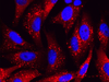
Chemical distribution of carbonates and phosphates in the middle crown of a bovine incisor control tooth (vertical section).
Experiments at the MIRAS beamline in ALBA
When analyzing the side effects of whitening treatments on enamel, most of the studies are focused on determining the changes in its mechanical properties, like surface hardness or roughness; however, these techniques are not able to provide information about the chemical changes taking place.
Fourier transformed infrared (FTIR) spectroscopy is a molecular vibrational technique that has been shown to be advantageous to investigate mineralized tissues like teeth, providing information on the chemical structure at the molecular scale. The addition of a microscope to FTIR microspectroscopy has led to the possibility of combining biochemical with spatial information. This is known as chemical imaging and has the potential to examine tissues at cellular resolution.
“Synchrotron-based Fourier transformed infrared microspectroscopy technique, available at the MIRAS beamline in the ALBA Synchrotron, can provide very precise chemical information in small areas, therefore it is a very suitable technique to study the changes induced by whitening treatments and to characterize the extent of its effects with precision.” Explains Ibraheem Yousef, scientist responsible of MIRAS beamline.
Article: Clara Babot-Marquillas, Maria-Jesús Sánchez-Martína, Jose Manuel Amigo, Ibraheem Yousef, Iris H.Valido, Roberto Boada, Manuel Valiente. Tooth whitening effects on dental enamel, oxidation or reduction? Comparison of physicochemical alterations in bovine enamel using Synchrotron-based Micro-FTIR. Dental Materials (2022). DOI: https://doi.org/10.1016/j.dental.2022.02.006

AtCDF3 gene induced greater production of sugars a...
Un estudio con datos de los últimos 35 años, ind...

Un equipo de investigadores de la Universidad Juli...

En nuestro post hablamos sobre este interesante tipo de célula del...
Horizon ha puesto en funcionamiento una nueva planta dedicada íntegra...