Researchers at the University of Barcelona (UB) and the Terrassa Health Consortium have found anatomical vulnerabilities of the brain reward system in obese people. By using magnetic resonance imaging techniques, the study reconstructed brain areas involved in the reward system and reported alterations in both the number of fibers and the integrity of neuronal connections. Results, published in the scientific journal Neuroimage, offer an in-depth analysis of obesity anatomical basis and provide new insights into the design of personalised therapies.
The study is led by María Ángeles Jurado, lecturer in the Department of Psychiatry and Clinical Psychobiology of the UB and researcher in the Institute for Brain, Cognition and Behaviour (IR3C), and Maite Garolera, head of the Neuropsychology Unit at the Terrassa Health Consortium, affiliated centre with the health campus of international excellence HUBc. The research team led by Martijn van den Heuvel, at the University Medical Center Utretch, collaborated in the study too.
An analysis of the reward system
With the prevalence of obesity increasing worldwide ―figures have doubled between 1980 and 2014―, understanding the processes leading to excessive eating behaviour becomes increasingly important. One of the hypotheses considered suggests that obesity is related to an abnormal structural wiring of the reward network to be associated with elevated food intake.
The reward system is a group of neural structures that are critically involved in mediating the effects of behaviour reinforcement. When the system is activated, it increases the probability of repeating the behaviour that produces its activation. The reward system has been particularly studied in the case of additions, but it is also the basis of food intake reinforcement. “The interest relies on describing and studying brain basis for food intake, particularly the anatomical basis of the reward system and its role in behaviour”, affirms Idoia Marqués, first author of the paper and researcher at the UB and the IR3C.
To achieve study’s aim, researchers used magnetic resonance techniques and selected sixty-three participants from 12 to 39 years old. Participants were divided into two groups: a first group composed by people with a body mass index equal to or higher than 30, which is the criteria established by the World Health Organization (WHO); and a second control group of people with a BMI between 18.5 and 25, which is considered as normal-weight for adults. To calculate the body mass index, the weight of a person in kilos is divided by his/her squared height in metres (kg/m²).
A sample of people with metabolically healthy obesity
One of the strengths of the study is the selection of a homogenous sample composed by metabolically healthy individuals, since inclusion criteria involved a history of any cardiovascular pathology, metabolic syndrome or psychological disorder. “Evidence supports that risk factors, for instance diabetes or cardiovascular diseases, produce structural and functional alterations in the brain. Therefore, if participants suffer any of these disorders, it is impossible to differentiate whether brain effects are related to obesity or to other factors. The sample enabled us to focus on obesity particular features”, points out María Ángeles Jurado.
Changes in brain connectivity
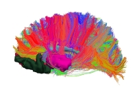
Researchers used magnetic resonance techniques to reconstruct the white matter pathways interconnecting the network of the reward system. Considering these data, they compared network wiring in terms of volume and integrity between both groups. “Results report evidence of an abnormal anatomical connectivity of the brain's reward system in obese subjects revealing lower levels of metrics of volume and integrity of anatomical pathways in the reward network”, explains Idoia Marqués.
“They are not big structural changes like in classical brain diseases; on the contrary, they are subtle changes. However, the existence of differences in the way intake is reinforced implies that the system that regulates eating behaviour is also different”, affirms María Ángeles Jurado.
Anatomical and functional differences
This is the first study in which researchers analyse reward network wiring. However, previous studies have already reported the existence of functional and anatomical brain differences in obese people. Findings provide evidence for cortical thickness in areas involved in cognitive processes and alterations in the reward processing of food stimuli.
The study may be useful to advance in obesity prevention and the development of personalized therapies. “Anomalous food intake is not always due to behaviour, but there is also a neuroanatomical substrate. This substrate, like other cultural and social aspects, must be considered in order to prevent obesity and design suitable treatments”, concludes María Ángeles Jurado.
Reference article:
Marqués-Iturria, I.; Scholtens, L. H.; Garolera, M.; Pueyo, R.; García-García, I.; González-Tartiere, P.; Segura, B.; Junqué, C.; Sender-Palacios, M. J.; Vernet-Vernet, M.; Sánchez-Garre, C.; De Reus, M. A.; Jurado, M. A.; Van den Heuvel, M. P. “Affected connectivity organization of the reward system structure in obesity”. Neuroimage, 2015, 111, p. 100-106. DOI: 10.1016/j.neuroimage.2015.02.012

AtCDF3 gene induced greater production of sugars a...

Un estudio con datos de los últimos 35 años, ind...

Un equipo de investigadores de la Universidad Juli...

En nuestro post hablamos sobre este interesante tipo de célula del...
Palobiofarma S.L. is pleased to announce the “last patient last visi...