Authors: D. Giosa, F. Casuscelli di Tocco, G. Raffa, C. Musolino, D. Lombardo, C. Saitta, R. Aiese Cigliano, W. Sanseverino, O. Romeo, G. Navarra, G. Raimondo, T. Pollicino
Institutions:
Publication: Digestive and Liver Disease
Date: February 2020
Full paper: https://www.dldjournalonline.com/article/S1590-8658(19)30964-8/abstract#articleInformation
Abstract:
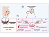
Infection with HBV is associated with a high risk of developing hepatocellular carcinoma (HCC). HBV may contribute to HCC development through both direct and indirect mechanisms. However, it remains undefined the relative contribution of virus-induced inflammation and the impact of viral integration. In this study, we evaluated HBV replication efficiency in tumor cells and analyzed the transcriptome of HBV-related HCCs to identify transcription of viral-human gene fusions from the genomic integration sites. Methods. HBV replicative and transcriptional activities were evaluated in tumor and non-tumor liver tissues from 5 patients with HBV-related HCC under NUC treatment using real-time PCR assays to quantitate total HBV DNA, cccDNA, and viral transcripts. Moreover, tumor and non-tumor liver tissues from 3 of the 5 patients, normal liver tissues from 3 individuals who underwent liver resection for hepatic hemangioma, and HepG2 cells were subjected to whole-transcriptome sequencing. RNA-seq was performed on an Illumina HiSeq 2500 platform. A total of 930 M reads were obtained. For chimeras detection a bioinformatics pipeline based on BWA, samtools, picard, bedtools, and in house scripts was used Results. Tumor and non-tumor liver tissues showed comparable mean amounts of total HBV DNA (355 ± 235 vs 298 ± 2150, copies/cell), cccDNA (0.26 ± 0.14 vs 0.16 ± 0.25 copies/cell), and transcripts (989 ± 937 vs 370 ± 256 copies/cell). Interestingly, transcriptome-sequencing data showed that whereas HepG2 did not express NTCP, tumor tissues expressed NTCP at levels comparable with those expressed both in non-tumor and in normal liver tissues. In addition, RNA-Seq allowed the identification of 531 integration breakpoints (14 in non-tumor and 517 in tumor tissues). Among genes target of viral integration we found an enrichment of genes implicated in redox, inflammatory, and metabolic processes as well as in signal transduction, cell cycle, and cell adhesion pathways. Conclusion. HBV can be integrated and efficiently replicate in tumor cells.

El equipo de investigadores observó cambios en el...

El gen AtCDF3 promueve una mayor producción de az...
Un estudio con datos de los últimos 35 años, ind...

En nuestro post hablamos sobre este interesante tipo de célula del si...
La revista ‘Nature Protocols’ selecciona esta técnica como “pro...