Investigators of the Vall d’Hebron Institute of Oncology’s (VHIO) Radiomics, Prostate Cancer Translational Research and Genitourinary Tumors Groups, have identified whole-body magnetic resonance imaging (WB-MRI) as a novel biomarker of systemic treatment response in metastatic castration resistant prostate cancer (mCRPC) with bone metastases. Published as a Research Letter in European Urology*, results show that WB-MRI can provide quantitative markers to monitor response and progression to therapy in bone metastases from prostate cancer.
Response evaluation criteria in solid tumors (RECIST) version 1.1 is currently the reference standard for assessing the efficacy of cancer therapies in people with solid tumors, especially in patients who are included in clinical trials. However, these guidelines have their limitations. They can only be used to evaluate a maximum of five tumors per patient and two per organ. Moreover, RECIST faces challenges in effectively assessing treatment response when cancer has spread to the bones, since bone metastases are not evaluable using this criterion.
“In patients with different metastatic sites, RECIST may fail to accurately assess response to therapy. Particularly in prostate cancer, since metastases most commonly develop in the bones. Current evaluation methods cannot capture treatment response of these metastases, representing a major challenge in patient management and drug development,” said Raquel Lopez-Perez, Head of VHIO’s Radiomics Group and co-corresponding author of this present study.
“We can use other clinical criteria for evaluating treatment response in these patients including pain, bone fracture, or prostate-specific antigen (PSA) levels in blood, but they are not always reliable indicators. We are therefore currently limited in predicting efficacy and deciding if a patient should continue with the same therapy or switch to another treatment modality,” observed Joaquin Mateo, a Medical Oncologist at the Vall d’Hebron University Hospital, Head of VHIO’s Prostate Translational Research Group, and co-corresponding author of this study.
The iPROMET multicenter clinical trial was designed to objectively quantify changes in apparent diffusion coefficient (ADC) that measures the magnitude of diffusion of water molecules within tissue, and other quantitative WB-MRI parameters. The investigators have identified various biomarkers of response including the balance between tumor cell density and fat content in bones.
Patients with metastatic prostate cancer and bone metastases are aged between 50 and 80 years, the majority of whom will have been previously treated with corticosteroids and therefore present accumulated fat in bone marrow.
“This fat disappears with the occurrence of bone metastasis and the density of tumor cells increases. Results of this study show that a higher cell density of bone metastases together with a low percentage of fat fraction are associated with an increased risk of disease progression,” explained Raquel Perez-Lopez.
Findings demonstrate that these biomarkers, in addition to circulating tumor DNA (ctDNA) analysis by liquid biopsy, can predict treatment response and progression of bone metastasis in mCRPC and therefore help guide treatment decision making in this patient population.
“This discovery may not have been relevant fifteen years ago. But now that we have various treatment modalities available, the ability to more accurately predict response to therapy will help us to select the optimal treatment strategy in a timely manner, thereby improving outcomes for our patients,” concluded Joaquin Mateo.
This research Project was supported by the Prostate Cancer Foundation, Instituto de Salud Carlos III, and La Marató de TV3 Foundation. Research led by Joaquin Mateo and Raquel Perez-Lopez at VHIO is also supported by the CRIS Cancer Foundation, the FERO Foundation. Spanish Association Against Cancer (AECC) and “la Caixa” Foundation.
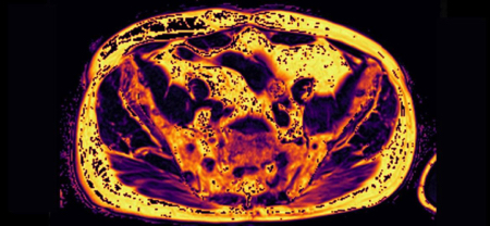
Image: Whole-body magnetic resonance imaging (MRI) of a 62-year-old patient with prostate cancer and bone metastases, who is responding to treatment. In the images in the first row, we can see how the accumulation of normal fat in the bone increases while, in the images in the second row, we can see how the density of tumor cells decreases.
*Garcia-Ruiz A, Macarro C, Zacchi F, Morales-Barrera R, Grussu F, Casanova-Salas I, Sanguedolce F, Gonzalez M, Cresta-Morgado P, de Albert M, Garcia-Bennett J, Marmolejo D, Planas J, Roche S, Mast R, Zatse C, Piulats JM, Herrera-Imbroda B, Regis L, Agundez L, Olmos D, Calvo N, Escobar M, Carles J, Mateo J, Perez-Lopez R. Whole-body Magnetic Resonance Imaging as a Treatment Response Biomarker in Castration-resistant Prostate Cancer with Bone Metastases: The iPROMET Clinical Trial. Eur Urol. 2024 Mar 14:S0302-2838(24)02133-X. https://doi.org/10.1016/j.eururo.2024.02.016

La mejor actitud que podemos adoptar es la de trat...

The research team observed changes in head circumf...

AtCDF3 gene induced greater production of sugars a...

En nuestro post hablamos sobre este interesante tipo de célula del...

Telum Therapeutics, a biotechnology company leveraging proprietary met...


