Researchers at the Centre for Genomic Regulation (CRG) have discovered how proteins work in tandem to regulate ‘treadmilling’, a mechanism used by the network of microtubules inside cells to ensure proper cell division. The findings are published today in the Journal of Cell Biology.
Microtubules are long tubes made of proteins that serve as infrastructure to connect different regions inside of a cell. Microtubules are also critical for cell division, where they are key components of the spindle, the structure which attaches itself to chromosomes and pulls them apart into each new cell.
For the spindle to function properly, cells rely on microtubules to ‘treadmill’. This involves one end of the microtubule (known as the minus end) to lose components while the other (the plus end) adds components. The effect is like that of a treadmill conveyor belt, where the microtubules appear to be moving continuously without changing their overall length.
Treadmilling is crucial for cell division. “The most likely theory is that treadmilling helps the cell regulate its attachments to chromosomes by maintaining tension. Because microtubules are often growing from their plus ends, this tension can be provided by constant shrinking from the minus ends,” explains Dr. Gil Henkin, co-first author of the study.
Despite the central role of treadmilling in cell biology, how the process is regulated has remained a mystery – till now. The authors of the study used various isolated proteins known to play a central role in microtubule biology, putting them together in a test tube and visualizing them using a microscope.
Three proteins were found to be critical for regulating treadmilling: KIF2A, a protein belonging to a larger family of proteins that dismantles microtubules, the γ-tubulin ring complex (γ-TuRC), a scaffold for microtubules to grow from, and spastin, an enzyme that acts like a scissor cutting microtubules.
“The family of proteins that dismantle microtubules usually nibble on microtubules at both ends. We were surprised to find that one member of this family – KIF2A – has a strong preference for minus ends. This specialization is exactly what researchers have been looking for to explain why microtubules treadmill in the spindle,” explains Dr. Thomas Surrey, senior author of the study and researcher at the Centre for Genomic Regulation.
Before KIF2A can nibble a minus end, it needs to overcome yTuRC, which acts like a safety cap. “The enzyme spastin is required to free microtubules from the safety cap so that KIF2A can do its job once microtubule plus ends have grown long enough,” explains Dr. Cláudia Brito, co-first author of the study.
The researchers found that the correct control of treadmilling requires the coordinated action of all three proteins. While the study does not directly translate into therapeutic avenues, it adds another piece to the intricate puzzle of cellular function and division. "Humans start as a single cell which must develop into many trillions of cells, all containing good copies of the genome. It's amazing and important that this process works extremely reliably, so we have added a small piece of the puzzle in understanding the overall mechanism,” concludes Dr. Henkin.
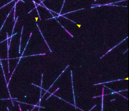
Image: KIF2A driving the treadmilling of microtubules

La mejor actitud que podemos adoptar es la de trat...

The research team observed changes in head circumf...

AtCDF3 gene induced greater production of sugars a...

En nuestro post hablamos sobre este interesante tipo de célula del...
New study finds an association between higher temperatures early in li...