Vall d’Hebron University Hospital achieves a double milestone in the field of lung transplants. For the first time ever, a lung has been transplanted using a minimally invasive technique that entails the use of robotic surgery. Also, a new access route has been created through which diseased lungs can be removed and the new lungs can be inserted. The new access route, which requires a mere eight-centimetre incision, was made in the lower part of the sternum, just above the diaphragm. This means it is no longer necessary to make a large opening by separating the ribs and opening up the thorax, which was the only available option until now. This pioneering procedure, which was performed on a 65 year-old man requiring a lung transplant due to pulmonary fibrosis, was carried out as part of a multidisciplinary intervention involving professionals from the Thoracic Surgery and Lung Transplants Department, the Anaesthesia, Resuscitation and Pain Management Department, the Cardiac Surgery Department and the Transplant Nursing Department.
Lung transplantation consists of replacing one or both diseased lungs with healthy ones. In general, this happens when there is a disease that involves severe and progressive chronic respiratory failure. Lung transplants began in 1981 in California. In Catalonia, this kind of procedure is carried out exclusively at the Vall d'Hebron University Hospital for both children and adults. Since the program began, more than 1,556 lung transplants have been carried out at Vall d'Hebron.
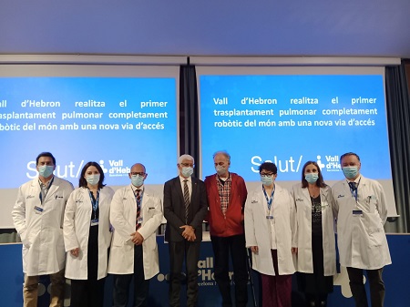
"Today we are proud to present a pioneering technique carried out by the Catalan Health-Care System that contributes to the clinical improvement of all patients internationally" says Manel Balcells, Minister of Health of Catalonia. “We present a new technique in lung surgery that represents an international and global advance. We do it together with Xavier, the first patient transplanted with robotic surgery and with a new, less invasive access route that allows a faster recovery”. Manel Balcells also states that Vall d'Hebron "is a reference center in lung transplantation for 10 million people: Catalonia, Aragon and the Balearic Islands. As a Public Health-Care system, we offer new techniques in global clinical practices that improve the well-being of all patients”.
“The main problem with opening up the thorax in lung transplant procedures is that it is a very aggressive approach which leads to a very delicate post-operative period”, explains Dr Albert Jauregui, head of the Thoracic Surgery and Lung Transplants Department at Vall d’Hebron University Hospital. In any transplant procedure, in order to prevent rejection of the new organ(s), medication must be administered that depresses the patient’s immune system for the rest of their life. This means that the risk of post-operative infection is always very high. In some cases, infection eventually occurs and the wound does not properly close (when both lungs are transplanted, the incision is about 30 centimetres-long, running from one side of the chest to the other). When the wound does not close due to the presence of an infection, it is necessary to re-operate to bring the infection under control. We must also note that patients in need of a lung transplant have chronic respiratory insufficiencies, and that simple actions such as going to the toilet can be exhausting for these individuals. Aggressive surgeries, such as traditional lung transplants, can therefore entail many negative consequences. But now, the paradigm has shifted: “This novel surgical technique allows us to cut a small section of skin, fat and muscle, leaving a wound that closes easily. Not only is this much safer than the traditional method, but for this first patient it has been virtually painless. This is a historic milestone which we believe will improve the lives of thousands of patients”, stated Dr Albert Jauregui.
The professionals of Vall d’Hebron University Hospital’s Thoracic Surgery and Lung Transplants Department had been planning to introduce robotic surgery to lung transplants for some time. This innovation had only been used once before, although in a less ambitious procedure, at the Cedars-Sinai Hospital in Los Angeles, USA. Last year, this American hospital used robotic surgery for the first time as part of a lung transplant when suturing the new lung to the patient’s airway and great vessels. However, the rest of the operation was performed in the traditional way and the lung was introduced through the ribs, as is customary.
“We at Vall d’Hebron had been thinking for some time about how we could make this very aggressive surgery less invasive. However, we were always faced with the same problem: we couldn’t work out a route to remove the diseased lung and insert the new one”, explained Dr Albert Jauregui. He added “finally, Dr Iñigo Royo Crespo, a specialist in the Thoracic Surgery and Lung Transplants Department, came up with the idea of exploring an access route used to operate on lung cancer and the thymus known as subxiphoid surgery.
The xiphoid is a small cartilaginous extension of the lower part of the sternum. Surgeons manually made an eight-centimetre incision in the skin below the xiphoid and above the diaphragm. In the open hole they placed a soft-tissue retractor: a simple plastic tool that serves to keep the incision open and clean during the operation to remove the diseased lungs and insert the new ones. The skin here is very elastic, so the eight centimetres are sufficient for the lungs to pass through. This differs from the incision made between two ribs, that is common in transplants, which is not elastic. From that point onwards, the operation was 100% robotic: four arms of the Da Vinci robot were inserted through four small holes (measuring 8 to 12 millimetres wide) made in different parts of the thorax. The thoracic surgeon sits at the console and moves the robot’s arms by means of four different control levers: one lever moves an arm that delicately separates the heart from the lung, so that it doesn’t hinder the removal or insertion of the lungs; two arms carry the surgical tools, such as scalpels and forceps; and the fourth arm incorporates a camera that allows the surgeon to have a 3D view of the inside of the body (remember that, until now, lung transplants were carried out by opening up the thorax so that the surgeon could see everything with the naked eye). The Da Vinci robot enables high-precision surgical interventions, as it offers excellent visibility and greater freedom of movement. Minimal, precise, and less invasive incisions can be made by means of this technology, which removes the risk factors of trembling, involuntary movements of surgeons and postural fatigue in long operations.
Once the patient’s lung was separated from the heart by the robotic arms, the diseased lung was removed through the subxiphoid opening. The new lung was then inserted through the same incision and attached to the body by the robotic arms. This is how the first fully robotic lung transplant was carried out at the Vall d’Hebron University Hospital, which could mark a real turning point in the history of lung transplants.
Lung transplants: a multidisciplinary task
A key speciality in all surgical operations is anaesthesia. As explained by Dr Maribel Rochera, head of the Anaesthesia, Resuscitation and Pain Management Department, these specialists “monitor the patient’s condition at all times, keeping them in the best possible condition throughout the operation. As this is a pioneering technique, we needed to combine our experience in both traditional transplants and robotic thoracic surgery, which involved a lot of teamwork”. Carme Vallès, supervisor of the Transplant Coordination Nursing Department, stated “this technique was completely new for all of us. However, we in the Nursing Department had been preparing for this moment for some time”. With this milestone, “the intensification of nursing care in the surgical process and the importance of the selection of the surgical nurse, perfusionist and anaesthetist to carry out the robotic operation is clear: a challenge that has been a success thanks to teamwork and professional consensus”.
When transplant patients leave the operating theatre they are always referred to the Intensive Care Unit, as this is where they receive the most appropriate care after such a complex operation. The first robotic lung transplant patient followed the same procedure. Dr Judit Sacanell, a lung transplant specialist in the Intensive Medicine Department, explained how “the role of the Intensive Medicine Department is key in the immediate postoperative period of transplant patients and the treatment of possible post-operation complications. We hope that this new technique will allow us to reduce the number of complications related to this type of surgical approach”. Finally, Dr Carles Bravo, medical director of the Hospital’s lung transplant programme, stated that “thanks to this important milestone, the lung transplant programme enters a new stage of minimally invasive surgery which offers multiple advantages for the lung transplant patient, which will improve the results of the lung transplant programme”.

La mejor actitud que podemos adoptar es la de trat...

The research team observed changes in head circumf...

AtCDF3 gene induced greater production of sugars a...

En nuestro post hablamos sobre este interesante tipo de célula del...

Telum Therapeutics, a biotechnology company leveraging proprietary met...


