The congenital heart diseases are a group of diseases that are characterized by the presence of structural abnormalities of the heart caused by defects in its formation during the embryonic period. In this context, in view of the long-term prognosis, myocardial remodelling plays a crucial role. In congenital cardiopathies, changes in the organization of the cells of the myocardium, in the mechanical properties, in the system of electrical conduction and in the blood supply to the myocardium, can lead to alterations in cardiac function, both before and after surgery.
To better understand and develop appropriate, sufficiently personalized treatment strategies, the microscopic organization of the cells that make up the heart muscle tissue and its integration at macroscopic level must be fully understood. The aim of a study published last October in the journal Circulation: Cardiovascular Imaging was to describe for the first time, in 3 dimensions and non-destructively the detailed remodelling of cardiac microstructure present in a human fetal heart with complex CHD (right isomerism).
The authors of the study, including its principal author Patricia Garcia-Cañadilla of the Department of Information and Communication Technologies (DTIC) at UPF, were led by Bart Bijnens, ICREA research professor of the DTIC and coordinator of the Sensing in Physiology and Biomedicine (PhySense) research group at UPF, and Andrew C. Cook, a researcher at University College London (UK), with the participation of researchers from the Barcelona Supercomputing Center (BSC) and research centres in Switzerland and Croatia.
Congenital disease has been studied using synchrotron light
Synchrotron X-ray phase-contrast imaging has enabled obtaining a detailed analysis of the organization of cardiomyocytes in 3D, which would not be obtainable with other imaging techniques. With this procedure the authors have been able to see clearly both microscopic and macroscopic changes in cardiac structure as well as in the electric conduction system specific to this complex congenital pathology. Thanks to this technique, disordered myocyte organization in the morphologically right ventricle myocardium has been observed for the first time. In addition, simulations of electrical activation conducted in cardiac tissue suggest the existence of an alteration in the synchronization of the right ventricle.
In their paper the authors state: We have shown the potential of X-ray phase-contrast imaging for studying cardiac microstructure in the developing human fetal heart at high resolution”, which provides a new vision and valuable information for future studies. This is the first study that shows alterations in the myocardium in midgestational congenital cardiopathies in human fetuses.
Reference work:
Patricia Garcia-Cañadilla, Hector Dejea, Anne Bonnin, Vedrana Balicevic, Sven Loncaric, Chong Zhang, Constantine Butakoff, Jazmin Aguado-Serra, Mariano Vázquez, Laurence H. Jackson, Daniel J. Stuckey, Cristoph Rau, Marc Stampanoni, Bart Bijnens, Andrew C. Cook (2018), "ComplexCongenital Heart Disease Associated With Disordered Myocardial Architecture in a Midtrimester Human Fetus", Circulation: Cardiovascular Imaging,15 october. 11: e007753.
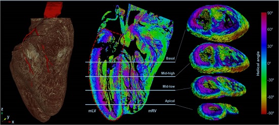
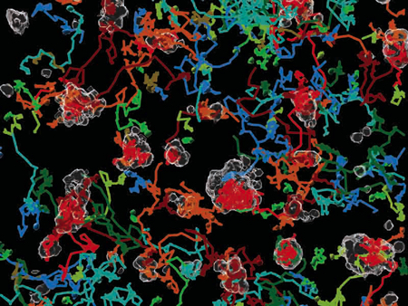
Image: Left-hand side: Volumetric model of the fetal heart showing details of heart anatomy along with the coronary arteries in red. Right-hand side: Results of the analysis of the local direction of the cells of the myocardium (the colour scale indicates the value of the helix angle (HA) of the cells in each voxel) along with the 3D representation of the vectors indicating the direction of the cells in 4 images at different apical-basal positions. Circulation: Cardiovascular Imaging. 2018; 11: e007753

The research team observed changes in head circumf...

AtCDF3 gene induced greater production of sugars a...
Un estudio con datos de los últimos 35 años, ind...

En nuestro post hablamos sobre este interesante tipo de célula del...
La revista ‘Nature Protocols’ selecciona esta técnica como “pro...