One of the risks associated with COVID-19 is the persistence of sequelae once the acute phase of the infection has been overcome. One of these is the inflammation that can remain in the tissue surrounding the alveoli of the lung and impair oxygen uptake. A study led by the Pneumology Service of the Vall d'Hebron University Hospital and the Pneumology group of the Vall d'Hebron Research Institute (VHIR) has made it possible to confirm histologically, by means of biopsies, those people who have suffered COVID-19 and who present this damage in the lungs and to differentiate them from those with healthy lungs. The results allow corticosteroid therapy to be administered only in those cases in which it is needed, thus avoiding side effects. The work, published in the journal CHEST , has been carried out in collaboration with the Anatomic Pathology and Radiodiagnostic Services of the same hospital and the CIBER of Respiratory Diseases (CIBERES).
Organized pneumonia is a type of diffuse interstitial lung disease characterized by a very important inflammation that can be derived from SARS-CoV-2 infection and that affects the tissue around the alveoli, prevents them from expanding properly during inhalation and, therefore, hinders lung function and oxygen uptake. The recommended treatment is the administration of corticosteroids, which can prevent pulmonary fibrosis from developing. It is important, however, to identify which patients really need the therapy, as it requires high doses of the drug for months and involves a high risk of side effects.
The paper by Vall d'Hebron researchers presents the results of the analysis of a group of patients at least 3 months after hospitalization for COVID-19. A thoracic CT scan was performed, as well as functional tests to assess lung function in 757 patients. If the results showed that there might be interstitial involvement in the lungs, biopsies were performed by bronchoscopy to reach, with a tube through the trachea, the internal part of the lungs. The biopsies were performed in 50 patients and the samples obtained made it possible to identify different levels of interstitial damage in the lungs.
Thanks to the biopsies, 30 people (60% of the total) were found to have severe lung damage and thus needed corticosteroid treatment, but there were 17 people (34%) who did not. "Thanks to performing the biopsies by bronchoscopy, we avoided the administration of corticosteroids to 17 people who, initially, according to the results of the CT and respiratory function tests, were recommended. It is important to confirm the lung damage to know if the treatment is recommended and thus avoid unnecessary side effects" , says Dr. Mario Culebras, specialist physician of the Pneumology Service of Vall d'Hebron University Hospital and researcher of the Pneumology group of VHIR. In the remaining 3 cases, although the biopsies indicated interstitial damage, it was decided not to administer the therapy due to an improvement in the clinical condition of the patients. In all cases the patients have been followed up to check their good evolution.
High-quality biopsies for understanding the sequelae of COVID-19
The biopsies were taken, as usual, using a cryoprobe, which has a metal tip that is rapidly frozen at -89ºC, allowing a small fragment of the lung to be obtained and analyzed in the laboratory. The Bronchoscopy Department of the Vall d'Hebron University Hospital, which has been using this technique for more than a decade, has the largest number of procedures performed in Catalonia. This method, which must be performed by specialized professionals, makes it possible to obtain high quality samples and, therefore, to make much more reliable diagnoses, without increasing the risk of complications.
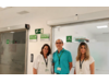
During the whole procedure, always performed with disposable material (including bronchoscope and cryoprobe), different professionals, such as pulmonologists, anesthesiologists and nurses, are involved. Nuria Neira and Laura Estepa, nurses from the Bronchoscopy Department of the Pneumology Service at Vall d'Hebron, explain that "patients with COVID-19 are especially fragile and need a lot of support, as some of them have spent a long time in the ICU. They are aware that tests such as these, despite being techniques that can be uncomfortable for them, are key to finding out what is wrong with them and receiving appropriate treatment to improve their clinical situation".
This was the first time that biopsy samples have been analyzed in living patients after suffering COVID-19 and, consequently, it has made it possible to evaluate the sequelae left in the lung tissue. Until now, this had only been done on autopsies of patients, most of whom had died during the acute phase of the disease. "Knowing this involvement in the lungs after severe SARS-CoV-2 pneumonia is important for initiating targeted therapy and reducing the sequelae" , concludes Dr. Culebras.