There are now about 150 million people in the world infected with hepatitis C, a disease that can lead to serious liver problems and is currently the leading cause of the need for liver transplants.
Researchers at the Centro Nacional de Biotecnología of the CSIC (CNB-CSIC) and the ALBA Synchrotron have now looked inside an infected cell in three dimensions and seen how the virus greatly distorts some of its structures. The study was recently published in the scientific journal ACS Nano.
"It's as if we had walked into the infected cell,” explains Pablo Gastaminza, principal author of the study. “We saw that the membranes of the endoplasmic reticulum as well as the mitochondria are extremely deformed."
The images from the study show how, as the infection advances, malformations of the endoplasmic reticulum increase and all the nearby mitochondria become distorted. Anomalies are also seen in the structures that allow communication between mitochondria and reticulum.
In addition, the authors observed that some of the antiviral drugs most commonly used to treat hepatitis C (daclatasvir + sofosbuvir) can reverse these structural alterations. “Using synchrotron light, we’ve seen for the first time how mitochondria and reticulum of infected cells recover their native conformation after only 7 days’ treatment with these drugs”, says Ana Joaquina Pérez-Berná, scientist at the ALBA Synchrotron and first author of the article.
A voyage to the interior of the infected cell
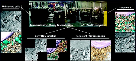
To generate this 3D map, the scientists used synchrotron light at ALBA, the complex of electron accelerators located in Cerdanyola del Vallès (Barcelona). With the MISTRAL beamline, they used a new technique called soft X-ray cryo-tomography (cryo-SXT). This method can obtain 3D images of the entire cell in its natural state, that is, without chemical pretreatment, cutting or drying the cell. “With this technique – only available in four places in the world (the ALBA Synchrotron, Bessy synchrotron in Germany, Diamond in the UK and ALS in the United States) – we can make a tomogram of the cell, similar to a conventional CT scan but with a million times more resolution,” says Pérez-Berná.
According to the scientists, the study shows that this new technique will be very useful for the study of the structural changes caused by pathological processes. "It could also be helpful in preclinical studies and for the development of new drugs, as it allows us to observe the effects of these compounds on different cell structures," says Gastaminza.
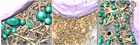
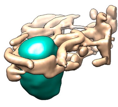
Figures: Interior of healthy cell, interior of a cell affected by the hepatitis C virus and interior of a cell after treatment with antiviral drugs. Violet cell nucleus, Green healthy mitochondria, beige healthy endoplasmic reticulum and yellow altered endoplasmic reticulum.
Ana Joaquina Pérez-Berná, María José Rodríguez, Francisco Javier Chichón, Martina Friederike Friesland, Andrea Sorrentino, Jose L Carrascosa, Eva Pereiro, and Pablo Gastaminza. Structural Changes In Cells Imaged by Soft X-Ray Cryo-Tomography During Hepatitis C Virus Infection. ACS Nano 2016 DOI: 10.1021/acsnano.6b01374
Image: Mitochondria and reticulum of healthy cell CNB-CSIC and ALBA Synchrotron

La mejor actitud que podemos adoptar es la de trat...

The research team observed changes in head circumf...

AtCDF3 gene induced greater production of sugars a...

En nuestro post hablamos sobre este interesante tipo de célula del...

Telum Therapeutics, a biotechnology company leveraging proprietary met...


