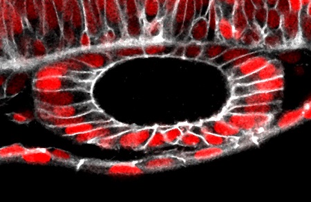
The study, published in Nature Communications on 16 June, shows for the first time that cells can exert forces that contribute to shaping cavities. The Developmental Biology Research Group, led by Berta Alsina, records the development of the ear in live zebrafish embryos. The research also involved Esteban Hoijman, first author of the article, and Davide Rubbini, both of the same research group, and Julien Colombelli, of the Institute of Biomedical Research of Barcelona.
The research has allowed ascertaining far more precisely than was previously possible how the cavity of the inner ear is formed, in this case taking the zebrafish embryo as a model. The results change the viewpoint that had existed to date on the formation of the cavities of organs.
The cavity formation process is decisive in the embryo, given their abundance in different organs (brain, heart, kidneys, intestines, blood vessels, etc.). Errors at this stage cause sometimes grave malformations, as is the case of spina bifida or polycystic kidney. In humans, hereditary deafness is the major cause of malformation in newborns, so understanding how this organ is formed is of vital importance.
Until now it was thought that the development of cavities (or lumen) of the organs was caused by a process similar to that of inflating a balloon: cells were the rubber and the external fluid would be the air. The results of this research show two changes to this appreciation. On the one hand, it has been seen that the fluid that fills the cavity not only comes from outside but also from inside the cells. On the other, it has been observed for the first time that mechanical forces also contribute to the expansion of the cavity and are caused by the change in shape when the cells divide.
In addition to its significant contribution to the knowledge of embryonic development, the relevance of research also lies in the fact of having used pioneering techniques that have allowed recording in vivo embryonic development of the ear and using laser microsurgery for the first time on internal three-dimensional epithelium. In the case of the zebrafish, which is transparent and whose ear is highly superficial, the area of the research was stained with fluorescence and viewed live. Laser microsurgery has been possible thanks to the use of a special microscope that has allowed the precise cutting inside the embryo of structures smaller than a cell without damaging them.
Understanding the mechanisms used by the organs to form cavities is also key for designing in vitro organ manufacturing strategies in regenerative medicine.
Reference work
Hoijman Esteban, Rubbini Davide, Colombelli Julien, Alsina Berta. "Mitotic cell Rounding and Epithelial Thinning Regulate lumen growth and shape". Nature Communications, June, 2015.

La mejor actitud que podemos adoptar es la de trat...

The research team observed changes in head circumf...

AtCDF3 gene induced greater production of sugars a...

En nuestro post hablamos sobre este interesante tipo de célula del...

Telum Therapeutics, a biotechnology company leveraging proprietary met...


