Detecting the parasite that causes malaria remains a challenge, particularly in resource-limited settings, as it often requires specialised laboratory equipment and trained personnel. To address this limitation, the Diagnostic Nanotools (DINA) group at the Vall d’Hebron Research Institute (VHIR) is working to facilitate malaria diagnosis through simple, low-cost technologies. Recently, the team developed a new paper-based device, less than 2 cm in length, that rapidly processes whole blood samples before analysis. The technology was developed in collaboration with the Institute for Bioengineering of Catalonia (IBEC) and Tufts University in the United States, and the results have been published in the journal Lab on a Chip.
Most cases of malaria are caused by infection with the parasite Plasmodium falciparum, which is not only the most prevalent species but also the most severe. The parasite enters the body through the bite of an infected mosquito and invades red blood cells (also known erythrocytes), where it multiplies until the cells rupture, releasing new parasites that infect additional erythrocytes. Effective diagnosis and treatment monitoring of malaria require the detection of the parasite itself or one of its components (biomarkers). To achieve this, blood samples are collected from patients and analysed for the presence of the parasite. “Before determining whether a blood sample contains the parasite, a preliminary step is required: the lysis, or disruption, of red blood cells. This releases Plasmodium antigens or genetic material contained within the cells and facilitates their detection”, explains Judit Prat, a predoctoral researcher in the Diagnostic Nanotools group at VHIR.
To perform this lysis step, specialised laboratory techniques are typically required. The work carried out by the DINA Group has focused on the design, fabrication and testing of paper-based devices capable of collecting and processing samples prior to analysis and that can be used in virtually any setting. The devices were developed in the laboratory of Dr Charles Mace, at Tufts University during a research stay by Judit Prat in 2024. The stay was supported by a mobility grant obtained within the framework of her industrial PhD fellowship, both funded by the Carlos III Health Institute.
A three-layer paper device to lyse red blood cells
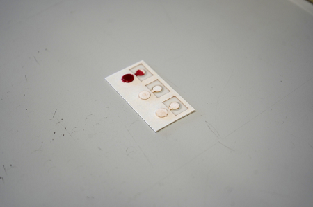
This paper-based device enables the lysis of red blood cells in small blood samples in less than ten minutes. It is a very simple system measuring approximately 2 cm in length and 1 cm in width, manufactured from paper using laser cutting.
The device consists of three layers, each with a specific function. The first layer receives the blood sample and contains a reagent that disrupts both the cells and the parasite. The second layer acts as a filter, separating waste products so that they do not interfere with the final measurement. Finally, the third layer consists of absorbent paper that collects the lysed sample containing the released cellular components.
As part of the study, the research team tested the device using Plasmodium-infected cell cultures generated by IBEC researchers and also with samples from real patients, comparing the results with conventional laboratory techniques. “We have shown that lysis performed with this system enables the detection of malaria parasite biomarkers with results comparable to those obtained using standard methods, but with a much simpler and lower-cost technology”, says Dr. Eva Baldrich, head of the Diagnostic Nanotools group at VHIR.
Once processed, the sample can either be analysed immediately or stored for later analysis. The study confirmed that samples can be maintained for approximately one week at room temperature, facilitating transportation and subsequent laboratory analysis when necessary.
A project to develop a new malaria detection technology
The VHIR team is currently developing rapid diagnostic systems to analyse samples obtained from finger-prick blood collection in malaria-endemic settings. Unlike commercially available rapid tests, the malaria test they are developing provides a quantitative assessment of the parasite in the patient's sample rather than simply detecting the presence or absence of infection. This makes it possible to determine the concentration of parasites in the blood (parasitaemia), helping to define treatment and patient follow-up more accurately. In addition, the technology is easy to manufacture, versatile and adaptable, and the VHIR team believes it could eventually be used to identify different types of microorganisms.
The development of these devices has been carried out within the framework of two projects: the European EuroNanoMed project (QUPID), funded in Spain by the Carlos III Health Institute and completed at the end of 2025; and the CATMAL project, which is supported by the Health Research call of the “la Caixa” Foundation.
Recently, the team travelled to Hospital Nossa Senhora da Paz in Cubal (Angola) to test both the rapid diagnostic assay and the lysis device, obtaining promising results. “Our ultimate goal is to integrate the lysis system and the detection assay into a single device in order to facilitate malaria diagnosis in settings without access to specialised laboratories”, concludes Dr. Baldrich.

La mejor actitud que podemos adoptar es la de trat...

The research team observed changes in head circumf...
AtCDF3 gene induced greater production of sugars a...
En nuestro post hablamos sobre este interesante tipo de célula del...
Un estudio liderado por la Universidad de Sevilla y el Centro de Inves...