CenSpark650: First-in-class dual-ligand fluorescent probe for selective imaging of centrioles and ciliary microtubules
Centrioles and cilia are essential organelles governing cell division, signalling, motility and immune function.
Despite their importance, studying their dynamics in live cells has remained technically restricted due to the lack of selective small-molecule probes.
Most existing approaches rely on genetic engineering or non-specific microtubule dyes, limiting their use in primary cells and non-model systems.
Key Challenges
Centrioles and cilia contain specialised microtubule architectures (triplets and doublets) that are absent from cytoplasmic microtubules.
However, no previously available small-molecule probe could selectively distinguish these structures in live cells.
As a result, researchers have been unable to:
Overview
Developed by Spirochrome, CenSpark650 is a cell-permeable small-molecule probe engineered to selectively recognise microtubule triplets and doublets found exclusively in centrioles and axonemes.
Its selectivity arises from a unique dual-binding mechanism targeting the spatially adjacent inner and outer microtubule binding sites of A- and B-tubules, a structural feature absent in cytoplasmic microtubules.
Key Benefits
Key Features
Typical Applications
Additional Resource
Read the full peer-reviewed study in Nature Chemical Biology. Pourroy, C., Hatzopoulos, G.N., Reymond, L. et al. Development of the fluorescent probe CenSpark for labeling centrioles and cilia. Nat Chem Biol (2026). https://doi.org/10.1038/s41589-026-02186-1.
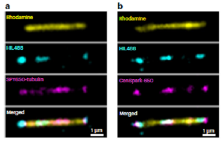
Imagen: CenSpark selectively binds microtubule doublets in vitro. a,b, Montage showing subtilisin-treated singlet microtubule (rhodamine, yellow) and doublet-like microtubule segments (HiL488, cyan), incubated with 2 nM SPY650-tubulin (a) or 2 nM CenSpark-650 (b) (both in magenta).
(Source: https://doi.org/10.1038/s41589-026-02186-1)

La mejor actitud que podemos adoptar es la de trat...

El equipo de investigadores observó cambios en el...

El gen AtCDF3 promueve una mayor producción de az...

En nuestro post hablamos sobre este interesante tipo de célula del si...
Desarrollan un nuevo enfoque basado en la fotofarmacología, que permi...