Bright, photostable, and cost-effective Acti-Stain™ probes for reproducible fluorescence microscopy.
Fluorescent phalloidins remain the gold standard for visualising filamentous actin (F-actin) in fixed cells and tissues. Their high specificity for polymerised actin makes them indispensable tools for studying cytoskeletal organisation, cell morphology, and signalling pathways in both basic and translational research.
Acti-Stain™ Phalloidin conjugates from Cytoskeleton Inc. offer a high-performance alternative to traditional FITC-labelled phalloidins. These probes combine exceptional brightness, superior photostability, and excellent value, making them ideally suited for multi-colour fluorescence microscopy and high-content imaging workflows.
Their partner, Cytoskeleton Inc. provides Acti-Stain™ phalloidins conjugated to three spectrally distinct fluorophores (488, 555, and 670). This enables seamless integration with commonly used nuclear and organelle stains across a wide range of microscope filter sets.
Independent comparisons with leading competing products demonstrate that Acti-Stain™ probes deliver comparable or superior image quality at a significantly lower cost, allowing laboratories to optimise both performance and budget, without compromising experimental reliability.
Key Advantages of Acti-Stain™ Phalloidins
Acti-Stain™ probes are:
Acti-Stain™ Product Overview
| Acti-Stain™ 488 Phalloidin | Acti-Stain™ 555 Phalloidin | Acti-Stain™ 670 Phalloidin |

|

|

|
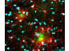
| Fig. 1: Fixed Swiss 3T3 cells stained for F-actin using Acti-Stain™ 488 (green, cat. no. 027PHDG1) and DAPI-labelled nuclei (blue). | Fig. 2: Fixed Swiss 3T3 cells with activated Rho. Nuclei stained with DAPI (blue); F-actin stained with Acti-Stain™ 555 (red, cat. no. 027PHDH1). | Fig. 3: Fixed Swiss 3T3 cells stained with Acti-Stain™ 670 (far-red F-actin, cat. no. 027PHDN1). |
Biochemical Performance of Fluorescent Phalloidins
To support informed reagent selection, Cytoskeleton Inc. extensively characterised Acti-Stain™ probes with respect to:
Table 1 compares Acti-Stain™ phalloidins directly with traditional FITC-phalloidin. The data demonstrate clear superiority in brightness and photostability, making Acti-Stain™ probes particularly suitable for demanding imaging applications.

Recent Publications Using Acti-Stain™

La mejor actitud que podemos adoptar es la de trat...

El equipo de investigadores observó cambios en el...

El gen AtCDF3 promueve una mayor producción de az...

En nuestro post hablamos sobre este interesante tipo de célula del si...
Desarrollan un nuevo enfoque basado en la fotofarmacología, que permi...