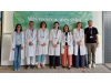
A team of researchers affiliated with IDIVAL, the Marqués de Valdecilla University Hospital, and the University of Cantabria has shown that a nanoplasmonic biosensor is capable of distinguishing tumor tissue from peritumoral brain tissue during brain metastasis surgery without the need for markers, stains, or external agents.
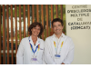
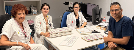
The work, published in Journal of Neuro-Oncology under the title A label-free nanoplasmonic biosensor for intraoperative discrimination of tumor margins in brain metastases surgery, also involved the Joan XXIII University Hospital in Tarragona and the General University Hospital of Elche. Among the authors are Víctor García-Milán and Laura Bauluz Olmedo as first authors, with Alfredo Franco and Carlos Velásquez coordinating the study.
The research addresses one of the main challenges in brain metastasis surgery: precisely determining where the tumor ends and healthy brain tissue begins. Although these lesions have traditionally been considered well defined, several studies have shown that microscopic infiltration may occur in the tissue surrounding the metastasis, an aspect that can influence local disease control.
To tackle this challenge, the researchers used a nanoplasmonic biosensor capable of detecting differences in the intrinsic optical properties of tissue. By analyzing the interaction of light with biological samples, the technology makes it possible to identify biophysical differences between tumor tissue and adjacent brain tissue without relying on dyes or contrast agents.
The study included 20 patients undergoing surgery for brain metastases, from whom paired samples of tumor and peritumoral tissue were collected intraoperatively following a standardized protocol. These samples were then analyzed using the biosensor and compared with histopathological examination, considered the reference method.
The results showed significant differences between the two types of tissue. In 85% of cases, tumor tissue presented higher refractive index values than peritumoral tissue, allowing the biosensor to discriminate between the samples.
This research continues a well-established line of work by the group in developing optical tools applied to oncological neurosurgery. In 2023, the team had already demonstrated the usefulness of this technology in differentiating glioblastoma from peritumoral tissue, and now expands its application to the field of brain metastases.
The researchers emphasize that this is a proof of concept and that the technology is not yet ready to replace anatomical pathology analysis or independently guide surgical decisions. Its potential lies in complementing other intraoperative tools and contributing to increasingly precise surgery.

La mejor actitud que podemos adoptar es la de trat...

The research team observed changes in head circumf...
AtCDF3 gene induced greater production of sugars a...
En nuestro post hablamos sobre este interesante tipo de célula del...
The direct analysis of human heart tissue from transplant recipients w...