Researchers at the Centro Nacional de Investigaciones Cardiovasculares Carlos III (CNIC) have developed a new computational method to study how the mammalian heart takes shape during the earliest stages of embryonic development.
The study, published in eLife, combines live microscopy of experimental models with advanced image analysis and machine learning. This approach allowed the researchers to reconstruct the movements and changes in shape that transform early cardiac tissue into the primitive heart tube.
The heart is the first organ to form and function in mammals. During its development, a flat group of precursor cells, known as the cardiac primordium, moves and changes shape to form a three-dimensional tube capable of beginning to pump blood.
Understanding this process could help explain how the heart develops normally and how alterations may lead to congenital heart defects. However, studying it is difficult because the tissue changes shape rapidly at the same time as the embryonic heart begins to beat.
“Live microscopy allows us to observe the developing heart, but converting these complex images into quantitative information has remained a major challenge. Our virtual map now makes it possible to follow how each region of the early cardiac tissue contributes to the primitive heart tube,” explains Dr. Morena Raiola, first author of the study and a researcher at the CNIC.
The researchers developed a computational method that tracks tissue movement directly from time-lapse microscopy images. They validated the method by comparing its results with the movements of individual cells tracked experimentally.
The team then used machine learning to align data from different embryos and combine them into a single model of cardiac development. This model shows where and when the tissue stretches, grows, or changes its direction of movement as the cardiac primordium becomes the linear heart tube.
A virtual map of heart development
One of the main results of the study is a virtual fate map that can track any point in the initial cardiac primordium and predict where it will be located in the primitive heart tube.
This map links the initial position of a cell or tissue region with the part of the heart tube to which it will later contribute. It therefore provides a new way to explore how the different regions of the embryonic heart are organized over time.
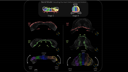
The analysis showed that neighboring cells generally move together in a coordinated way. However, not all parts of the tissue deform in the same manner. Instead, the cardiac primordium is divided into distinct regions, each with its own pattern of movement and growth. These different but coordinated patterns transform the initially bilateral cardiac primordium into a longitudinal, three-dimensional heart tube.
The study also provides new information about the formation of the primitive ventricular chamber. In the region that will become the outer curve of the heart tube, the tissue expands into a shape resembling half a barrel. This expansion is limited at both ends by two belt-like regions: one at the arterial pole and the other at the venous pole. These regions guide tissue growth and help give the developing ventricle its characteristic shape.
“The main advance is that we can now describe heart tube formation quantitatively and show that the primitive heart is shaped by distinct tissue regions following specific but precisely coordinated patterns of deformation,” explains Dr. Miguel Torres, head of the Genetic Control of Organ Development and Regeneration Group at the CNIC and senior author of the study.
A method for studying other organs
The new computational method could also be used to study other organs and tissues that undergo complex three-dimensional changes during embryonic development.
This fundamental research provides essential knowledge about how the heart forms normally, laying the groundwork for future studies into how alterations in this process may contribute to congenital heart defects.
The findings also provide a new framework for understanding the physical and cellular mechanisms that shape the embryonic heart. In the longer term, this knowledge could support research into congenital heart disease and tissue-engineering strategies.
The editors of eLife describe the study as an important advance in understanding tissue deformation and growth during the earliest stages of mammalian heart development. They also highlight the originality of the computational method and its ability to extract new biological information from live microscopy data.
The study received funding from the European Union’s Horizon 2020 Marie Skłodowska-Curie programme (H2020-MSCA-ITN-2016-722427); the Spanish State Research Agency through projects PGC2018-096486-B-I00 and PID2022-140058NB-C31; the Comunidad de Madrid through CARDIOBOOST-CM (P2022/BMD-7245); and the European Research Council through the REACTIVA Advanced Grant (101142005).
The research also received support from the CNIC Microscopy and Dynamic Imaging Unit, supported by FEDER ‘Una manera de hacer Europa’ and part of the ReDIB ICTS infrastructure TRIMA@CNIC and co-funded by the European Regional Development Fund.

La mejor actitud que podemos adoptar es la de trat...

The research team observed changes in head circumf...
AtCDF3 gene induced greater production of sugars a...
En nuestro post hablamos sobre este interesante tipo de célula del...
The direct analysis of human heart tissue from transplant recipients w...