International research in which the UPV/EHU’s Magnetism and Magnetic Materials group is collaborating has succeeded in individually characterising the nanomagnets contained in these organisms.
Imagine a tiny vehicle, a nanocar (one million times smaller than a millimetre), equipped with a magnetic structure that allows it to be controlled and steered by means of magnetic fields. Let's imagine that we can insert this car into the human body and send it to the exact spot where a drug needs to be released or cancer cells need to be eliminated. Numerous scientists across the world are working on this bold idea, including the multidisciplinary Magnetism and Magnetic Materials group (GMMMT) at the UPV/EHU-University of the Basque Country. This team is involved in research that has been published in the journal ACS Nano and is taking a new step towards turning the idea into reality.
Specifically, this group, led by the Faculty of Science and Technology lecturer Maria Luisa Fernández-Gubieda, is exploring the use of magnetic bacteria, known as magnetotactic bacteria, in the fight against cancer. These microorganisms have the amazing ability to form magnetic iron oxide nanoparticles inside their cells. The particles measuring about 50 nanometres across (100 times smaller than blood cells) are arranged inside the bacterium in the form of a chain which acts like a magnetic compass and guides the bacterium in its entirety in the direction specified by a magnetic field. The idea would be to use them to treat cancer by means of magnetic hyperthermia or to release drugs: the bacteria would be directed to the site of the tumour, and heated by external fields enabling them to burn cancer cells and/or release drugs by heat or another external stimulus.
Now, in collaboration with a team from the Helmholtz-Zentrum Berlin led by Sergio Valencia, they have been able to explore the magnetic properties of these bacteria in more detail. The degree of success of all the possible applications depends on the magnetic properties of these bacteria, and in particular, on each of the nanomagnets that form their chains. However, the magnetic signal from a single particle is so weak that, until now, it has been necessary to study the response of averages of hundreds or thousands of nanoparticles to obtain meaningful results.
Having only these averaged values restricted the design of customised nanomagnet applications. And this is what has now changed. Physicist Lourdes Marcano, a GMMMT member, has developed a new method. "We can now obtain precise information about the magnetic properties of several individual nanomagnets simultaneously," she said.
Magnetic anisotropy
Indeed, the new method makes it possible to measure the magnetic properties of individual magnetic nanostructures, even when they are inside biological entities. In particular, thanks to the magnetic images obtained in the X-ray transmission microscope of the BESSY II synchrotron (Helmholtz-Zentrum Berlin), and with the help of theoretical simulations, they have obtained precise information on the magnetic anisotropy of each nanoparticle within the microscope's field of view. Magnetic anisotropy describes how a magnetic nanoparticle reacts to external magnetic fields applied in an arbitrary direction. It is therefore an important parameter for controlling and directing magnetic nanoparticles.
At the moment, obtaining magnetic images of magnetic nanoparticles inside a biological cell with sufficient resolution is only possible in large synchrotron radiation facilities, such as the ones at the Helmholtz-Zentrum Berlin. "However, in the future, with the development of compact plasma X-ray sources, this method could become a standard laboratory technique," said Sergio Valencia.
"The bacterium is an excellent magnetic model that helps us to understand the behaviour of magnetic nanoparticles and to develop models that transcend to other systems," explained Mª Luisa Fernández-Gubieda. Her group is currently working on controlling the mobility of the bacteria by means of external magnetic fields to direct them to the tumour and activate them, also by means of magnetic fields, so that they perform the desired function.
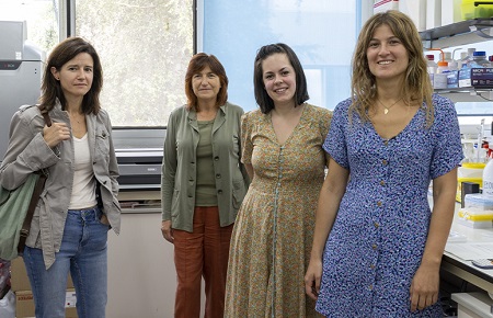
Image: From left to right, the researchers of the Magnetism and Materials Group Ana García Prieto, Maria Luisa Fernández Gubieda, Lourdes Marcano y Lucía Gandarias | Photo: Unai Zorriketa. Communication office. UPV/EHU
Bibliographic reference

La mejor actitud que podemos adoptar es la de trat...

The research team observed changes in head circumf...

AtCDF3 gene induced greater production of sugars a...

En nuestro post hablamos sobre este interesante tipo de célula del...

Telum Therapeutics, a biotechnology company leveraging proprietary met...


