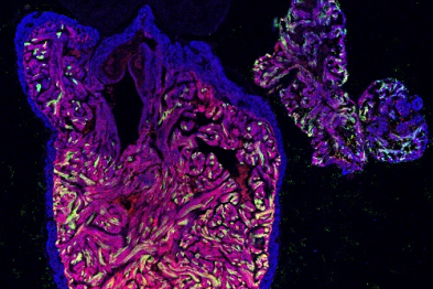
Some animals, including the zebrafish, have a high capacity to regenerate tissues, allowing them to recovery fully after cardiac injury. During this process, the heart muscle cells divide to replace the damaged tissue. However, there has been uncertainty about whether all cells contribute equally to the reconstruction of the heart wall. Now, a team of scientists led by Nadia Mercader at the Centro Nacional de Investigaciones Cardiovasculares Carlos III (CNIC) and the University of Bern (Switzerland), working with collaborators at the University of Zurich (Switzerland), have discovered a high level of plasticity among the cells of the zebrafish heart muscle. The study is published in Nature Communications.
After a heart attack, the human heart loses millions of cardiomyocytes, the cells that form the muscle wall. In contrast, other animal species have a high regenerative capacity, enabling them to replace the injured myocardium with new cardiomyocytes. One such species is the zebrafish (Danio rerio). According to first author Héctor Sánchez-Iranzo, the zebrafish “is a widely used model system in cardiovascular research into the mechanisms controlling regeneration, and an inspiration for attempts to develop future regenerative therapies.”
The ability of the zebrafish heart to reestablish its function after injury depends on the capacity of its cardiomyocytes to divide and repopulate the infarcted area, thus eliminating the damaged tissue. Unfortunately, the hearts of most animals, including humans, are unable to activate this process, and therefore after an infarction the human heart cannot regenerate the lost muscle, which is replaced by non-functional scar tissue.
Cell plasticity
Before the new study, scientists did not know if all cardiomyocytes in the zebrafish heart shared the same regenerative ability or if they were equally able to regenerate all zones of the heart muscle. Cell plasticity, the ability of cells to convert themselves into another cell type, is frequently observed during embryonic development, but has never before been reported during tissue regeneration in an adult organism.
In the study, which received funding from the European Research Council (ERC Starting Grant 2013 337703 zebraHeart), the authors investigated two types of cardiomyocyte, one localized in the innermost heart regions, the trabeculae, and the other in the exterior heart wall. Scientists had presumed that during regeneration each cardiomyoctye population would give rise only to the same specialized cell type. But the CNIC study shows that cardiomyocytes from the trabeculae can contribute to the regeneration of the external heart wall. The researchers conclude that their results “reveal a high level of plasticity among zebrafish cardiomyocytes and that there is more than one way to rebuild a damaged heart.”

La mejor actitud que podemos adoptar es la de trat...

The research team observed changes in head circumf...

AtCDF3 gene induced greater production of sugars a...

En nuestro post hablamos sobre este interesante tipo de célula del...

New study finds an association between higher temperatures early in li...