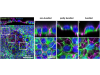
A study conducted by the University of Barcelona provides new evidence on the role of the glymphatic system, responsible for clearing waste from the brain, in neurodegenerative diseases. The study shows that patients with conditions such as Alzheimer’s disease, frontotemporal lobar degeneration and amyotrophic lateral sclerosis (ALS) exhibit a higher accumulation of wasteosomes, small structures that act as waste containers, in key regions of the waste-clearance system. These results suggest that the brain’s waste clearance system may be functioning poorly in these patients, which could lead to the accumulation of these structures and contribute to the progression of the disease.
According to researchers, understanding how the brain removes waste could be key to better understanding neurodegenerative diseases and, in the future, developing new prevention and treatment strategies.
The study, published in the journal Acta Neuropathologica Communications, was led by Professor Jordi Vilaplana, from the UB’s Faculty of Pharmacy and Food Science, the Institute of Neurosciences (UBneuro) and the CIBER Area for Neurodegenerative Diseases (CIBERNED), together with Laura Molina-Porcel, a researcher at the Neurological Tissue Bank Biobank of the Hospital Clínic de Barcelona – August Pi i Sunyer Institute of Biomedical Research (Biobank HCB-IDIBAPS). Marta Riba and Raquel Alsina (UB-UBneuro-CIBERNED) are the first authors of a study in which researchers from the Universitat Politècnica de Catalunya and Qilimanjaro Quantum Tech also participated.
Brain waste in key regions
The researchers analysed brain samples from 185 donors, including people with Alzheimer’s disease, amyotrophic lateral sclerosis and frontotemporal lobar degeneration, as well as individuals without a neurodegenerative disease. The aim was to determine whether patients exhibit differences in the number of wasteosomes, also known as corpora amylacea, and where these structures appear.
The results of the analysis show that wasteosomes repeatedly accumulate in specific regions of patients’ brains, specifically in areas associated with the drainage pathways of the glymphatic system, and that their number is significantly higher in the brains of diseased donors. According to the researchers, this distribution reinforces the hypothesis that wasteosomes may act as indicators of chronic glymphatic system insufficiency, that is, that the system responsible for clearing waste from the brain does not function correctly over a prolonged period of time. This distribution is also independent of the distribution of the pathological proteins characteristic of each disease. “This finding is particularly relevant because it indicates that wasteosomes are not a local consequence of deposits of altered proteins, but rather reflect a common mechanism shared by different neurodegenerative diseases,” the researchers explain.
In this context, the glymphatic system emerges as a potentially key element in understanding this shared mechanism. "When this system functions inefficiently, the brain’s ability to eliminate altered proteins and other waste products is reduced, which could promote the accumulation of toxic substances and contribute both to the onset and progression of neurodegenerative diseases,” the experts note.
A UB study identifies a common pattern of brain waste accumulation in patients with Alzheimer’s disease, amyotrophic lateral sclerosis (ALS) and frontotemporal lobar degeneration.
A window into the brain’s clearing System
One of the main implications of the study is that wasteosomes could become an indirect biomarker of chronic glymphatic insufficiency. “At present, we have very few indicators that allow us to assess the functional state of the glymphatic system, especially when it comes to alterations that have developed over years,” the researchers say.
Furthermore, if future studies confirm this relationship between wasteosomes and glymphatic insufficiency, they could also contribute to the development of new biomarkers to help identify individuals at greater risk of developing neurodegenerative diseases or to monitor their progression.
Along the same lines, the results reinforce the importance of investigating strategies aimed at preserving or restoring the functioning of the glymphatic system. “If its alteration contributes to the development of various neurodegenerative diseases, as numerous studies indicate, improving its function could become a new way to reduce the accumulation of brain waste and slow the progression of the disease,” the researchers note.
To move towards these potential diagnostic and therapeutic applications, the next step for the UB research team is to obtain direct evidence linking the presence of these structures to a functional alteration of the brain’s waste clearance system. To achieve this, the researchers note that experimental studies combining assessment of the glymphatic system’s function with analysis of wasteosomes will be required, both in animal models and, where possible, in human studies.
“It will also be important to extend this type of analysis to other neurodegenerative diseases and to develop tools that allow the detection of wasteosomes, or the changes they produce in the brain, using non-invasive imaging techniques,” the researchers conclude.

La mejor actitud que podemos adoptar es la de trat...

The research team observed changes in head circumf...
AtCDF3 gene induced greater production of sugars a...
En nuestro post hablamos sobre este interesante tipo de célula del...
El trabajo del Centro de Investigación del Cáncer (CSIC-USAL) demues...