There are epilepsy patients due to sclerosis of the hippocampus who, after their removal, continue to suffer episodes of crisis. In this study, they define a method of measuring hippocampal connectivity, in the hemisphere not damaged by epilepsy, which could help predict the efficiency of surgery before the intervention, and thus better define the risks and advantages that it may entail.
Most cases of epilepsy can be treated with drugs. However, there are certain occasions when that medication is not enough that surgery becomes necessary. This is the case of epilepsies caused by hippocampus sclerosis. In this type of epilepsy, the damaged hippocampus is removed, which, in most cases, leads to a significant improvement in patient condition. Even so, 30% of patients improve, but crisis episodes remain after surgery.
In this study, conducted at the Bellvitge Biomedical Research Institute (IDIBELL) and the Bellvitge University Hospital (HUB) and Barcelona University (UB) and published in a European journal of epilepsy, Seixure, they define certain parameters that could predict whether patients, with this type of epilepsy undergoing surgery, would present a good postoperative prognosis. To do this, before surgery, they compared different types of MRI imaging from patients who had completely abolished crisis episodes, "cured" patients, and patients who had not, "uncured" patients.
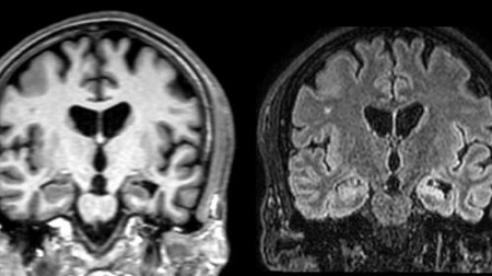
Most studies of this type of epilepsy focus on the analysis of the damaged region, the sclerotic hippocampus. However, Dr. Estela Cámara researcher from IDIBELL and Medicine and Health Science School of UB, principal investigator, points out that, in the study, they have focused on the examination of the other hemisphere hippocampus, the one that has not been damaged. This has allowed them to define hippocampus subregions through a high-resolution MRI, and subsequently determine connectivity in each subregion through diffusion MRI, or in other words, define the integrity of the wiring. After studying patients for 5 years, they observed that those with the persistent crisis have a more damaged and disorganized hippocampus than those in which the crisis had disappeared.
Dr. Jacint Sala, the first author of the study, explains that "the brain is a highly organized structure, since neurons are organized into large beams or columns, and we can characterize the configuration of these beams and establish a connectivity measurement through diffusion images". In diffusion resonance, we measure the movement of water between these large beams, when the structure and connectivity in a region is lost, water flows more easily in all directions and loses directionality. In this study, the quantification of subregions connectivity in the non-operated hippocampus gives a new tool to predict the effectiveness of sclerotic hippocampal extirpation in the disappearance of patient's crisis.
These results indicate that those patients who do not overcome the episodes of crisis after the operation, could present more extensive damage to the brain, the disease could have evolved to the point of not being able to be reversed by surgery.

La mejor actitud que podemos adoptar es la de trat...

The research team observed changes in head circumf...

AtCDF3 gene induced greater production of sugars a...

En nuestro post hablamos sobre este interesante tipo de célula del...

New study finds an association between higher temperatures early in li...