Cancer begins and develops in the realm of genes and proteins. It is a world inscrutable to the naked eye, but accessible through increasingly precise imaging techniques. Such as immuno-PET, which allows us to observe, live, in real time and on a molecular scale what is happening as soon as the disease appears.
This new medical imaging technique has already proved its potential for early diagnosis and treatment of lung, hematological and breast tumours, as Francisca Mulero, head of the Molecular Imaging Core Unit at the Spanish National Cancer Research Centre (CNIO), explains in an editorial in the journal Frontiers in Medicine.
Immuno-PET is an “innovative technique capable of revolutionising disease diagnosis, therapeutic decisions and patient outcomes,” says Mulero.
Immuno-PET is based on the PET (Positron Emission Tomography) scanner. In PET, a small amount of a radioactive chemical, or radiopharmaceutical, is inoculated into the body. The most common radiopharmaceutical contains glucose, the foodstuff for cells; when it accumulates on spots where cells are consuming more energy, it starts emitting detectable radiation (the image lights up). Visualizing those areas with a more active metabolism can help identify cancer cells, which consume glucose faster than healthy ones.
Antibodies as guides for better accuracy
Immuno-PET images show higher precision than conventional PET because the radiopharmaceutical is much more specific in pinpointing events of medical interest. This is achieved by attaching to the radioactive chemical a kind of antibodies designed according to the molecules or processes to be detected.
Antibodies are the proteins that allow the body to recognise particular enemies. In nature, when we are attacked by a foreign agent, the immune system generates antibodies specifically designed to deal with it: they have such a three-dimensional shape that they fit precisely into other proteins only characteristic to the invading enemy, like a key in a lock.
In recent decades, science has learned how to make in the lab customized antibodies, that is, antibodies shaped to recognize molecules of interest. These antibodies are the basis of targeted therapies, which aim to reduce side effects and increase treatment effectiveness. If, for example, an antibody is coupled to a drug, the drug will specifically reach the cells it should act upon, precisely those with the proteins the antibody can fit into, according to its design.
Immuno-PET in lung, hematological and breast tumours
In immuno-PET, antibodies guide the radiopharmaceutical to the processes or tissues to be studied. This makes it possible to observe in real time and in living organisms the molecular changes related to the onset –or very early stages– of diseases, their progression and their response to drugs.
One of the most promising areas of application is oncology, where its potential has been demonstrated for the detection and monitoring of the disease, as well as for checking the response to treatment in lung, hematological and breast tumours.
Mulero’s review includes, among others, work led by Anis Krache of the Centre de Recherche du Cancer de Toulouse (CTRT-INSERM, France), which showed how an immunotherapy drug was distributed in lung tumour tissue.
Nanobodies’ to visualise breast cancer metastases
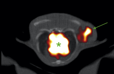
Mulero herself, in collaboration with Jorge L. Martínez Torrecuadrada, head of the Protein Production Unit at CNIO, and others, has used immuno-PET to observe metastases of the most aggressive type of breast cancer, triple-negative breast cancer.
In their study, they used nanobodies, antibodies from camelids (camels, llamas and alpacas) and sharks, which are ten times smaller than human antibodies. The CNIO researchers found that it was easier for nanobodies to reach their target. In addition, their smaller size allows the organism to eliminate the radiopharmaceutical sooner.
In any case, Mulero says that limitations or problems such as “tracer design, radiochemistry and translation to clinical practice” still need to be elucidated. She stresses that “more research and collaboration between scientists, clinicians and industry representatives is needed” to develop the full potential of the technique.
Reference article: Mulero Francisca, “Editorial: ImmunoPET imaging in disease diagnosis and therapy assessment”. Frontiers in Medicine. 2023, vol. 10.
https://doi.org/10.3389/fmed.2023.1231525
Image: ImmunoPET of a triple-negative breast cancer model using labelled nanobodies. The arrow points to the tumour and the asterisk to the heart. / Francisca Mulero

La mejor actitud que podemos adoptar es la de trat...

The research team observed changes in head circumf...

AtCDF3 gene induced greater production of sugars a...

En nuestro post hablamos sobre este interesante tipo de célula del...

Telum Therapeutics, a biotechnology company leveraging proprietary met...


