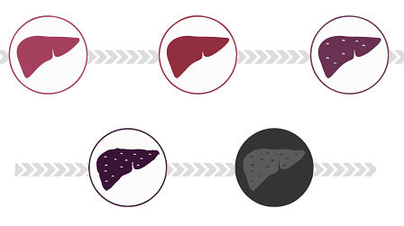
Non-alcoholic steatohepatitis (NASH) is the advanced form of non-alcoholic fatty liver disease (NAFLD) characterized by an accumulation of fat in the liver cells that causes inflammation, ballooning of the liver cells, and hepatic fibrosis. The inflammatory process, if severe, can cause significant damage and eventually lead to cirrhosis, which involves scarring of the liver tissue in the form of chronic and fatal injury. In addition, extensive fibrosis can also lead to hepatocellular carcinoma (HCC), which is also life-threatening.
Although the pathway for NASH development is still to be elucidated, many proposed mechanisms account for the increased fat accumulation in the liver cells, as well as many comorbidities. Particularly, obesity, insulin resistance (type 2 diabetes mellitus), and hyperlipidemia are some of the common metabolic comorbidities. Moreover, NASH has been linked to an increased risk for developing cardiovascular diseases and chronic kidney disease (Kaps et al., 2020; Peng et al., 2020). It is known that with insulin resistance, the buildup of adipose tissue on the liver releases free fatty acids into the hepatic tissue that can be stored as triglycerides in liver cells and cause steatosis or be metabolized into lipotoxic lipids. When these molecules build up in hepatocytes, they can cause stress responses within the cell which lead to inflammation and potential cell damage. Thus, when these cells become injured, the body activates the recruitment of inflammatory cells and the release of cytokines which can stimulate stress in surrounding cells or trigger hepatic stellate cells which cause the deposition of collagen fibers leading to fibrosis (Peng et al., 2020).
In these models, chemical and dietary alterations were used and as a result, it was detected that western diet (consisted of high cholesterol, high fructose, and high fat) in mice led to the developed of fibrosis and inflammation present in stage 3 of fibrosis at only 12 weeks and further developed HCC by 24 weeks. Data obtained using these models mimic quite accurately the same characteristics and progression of human NASH and could serve as a valuable tool in the drug testing process (Tsuchida et al., 2018).
On the other hand, it should be emphasized that in-vitro cell culture in 2D monoculture and 3D hepatic organoids and spheroids have been able to replicate key physiological symptoms of NASH. Induction of 2D hepatocyte culture with FFA has shown the same triglyceride accumulation in the cells that is responsible for the signaling cascade that leads to inflammation and fibrosis/cell death, although this model lacks the interaction of hepatocytes with non-parenchymal cells in co-culture which is necessary for the inflammatory process. However, 3D models can react to FFA in the way that causes hepatocyte ballooning and fibrosis that leads to inflammation and collagen production (Soret et al., 2021). Overall, both in-vitro models showed the capability to reproduce the pathophysiological features associated with NASH outside a living organism making them a key alternative for drug development, much cheaper than in-vivo models.
To conclude, NASH is a growing epidemic alongside obesity and type 2 diabetes mellitus with potentially life-threatening complications and comorbidities. Without a clear pathogenesis or treatment plan, the continuous damage of NASH on liver cells and tissues is mostly irreversible. However, essential in-vitro models have been developed that have shown to help us understand NASH disease progression, and thus allow early identification and treatment of this disease to avoid liver failure or the need for a liver transplant in the future.
References used:
Estes, C., Razavi, H., Loomba, R., Younossi, Z., & Sanyal, A. J. (2018). Modeling the epidemic of nonalcoholic fatty liver disease demonstrates an exponential increase in burden of disease. Hepatology (Baltimore, Md.), 67(1), 123. https://doi.org/10.1002/HEP.29466
Kaps, L., Labenz, C., Galle, P. R., Weinmann-Menke, J., Kostev, K., & Schattenberg, J. M. (2020). Non-alcoholic fatty liver disease increases the risk of incident chronic kidney disease. United European Gastroenterology Journal, 8(8), 942. https://doi.org/10.1177/2050640620944098
Peng, C., Stewart, A. G., Woodman, O. L., Ritchie, R. H., & Qin, C. X. (2020). Non-Alcoholic Steatohepatitis: A Review of Its Mechanism, Models and Medical Treatments. Frontiers in Pharmacology, 11. https://doi.org/10.3389/FPHAR.2020.603926
Soret, P.-A., Magusto, J., Housset, C., & Gautheron, J. (2021). In Vitro and In Vivo Models of Non-Alcoholic Fatty Liver Disease: A Critical Appraisal. Journal of Clinical Medicine, 10(1), 36. https://doi.org/10.3390/JCM10010036
Tsuchida, T., Lee, Y. A., Fujiwara, N., Ybanez, M., Allen, B., Martins, S., Fiel, M. I., Goossens, N., Chou, H.-I., Hoshida, Y., & Friedman, S. L. (2018). A Simple Diet- and Chemical-Induced Murine NASH Model with Rapid Progression of Steatohepatitis, Fibrosis and Liver Cancer. Journal of Hepatology, 69(2), 385. https://doi.org/10.1016/J.JHEP.2018.03.011

La mejor actitud que podemos adoptar es la de trat...

The research team observed changes in head circumf...

AtCDF3 gene induced greater production of sugars a...

En nuestro post hablamos sobre este interesante tipo de célula del...

New study finds an association between higher temperatures early in li...