A study conducted by the INCLIVA Health Research Institute, the Clinical University Hospital of València, the University of Valencia (UV), the Universitat Politècnica de València (UPV), the Cardiac Imaging Unit of Ascires Biomédico Group, and the Ascires-UPV Joint Research Unit provides significant evidence in the field of advanced cardiac imaging and clinical cardiology. It shows that the damage resulting from a myocardial infarction is not confined solely to the affected area, but may also functionally extend to remote regions of the ventricle. Identifying this dysfunction could have important implications, including improved prognostic assessment of patients, earlier detection of cardiac deterioration before symptoms or more evident changes appear, and the implementation of earlier and more personalised therapeutic strategies.
The study focuses on the consequences of myocardial infarction, one of the main manifestations of ischaemic heart disease. Globally, cardiovascular diseases remain the leading cause of death. According to the World Health Organisation, they account for nearly 18 million deaths per year, with myocardial infarction representing a very significant proportion. In Spain and other developed countries, survival rates following a heart attack have improved considerably thanks to advances in acute treatment. However, this has increased the number of patients living with chronic sequelae, such as ventricular dysfunction and the risk of heart failure. In this context, techniques such as cardiac magnetic resonance imaging with myocardial strain analysis are emerging as key tools for moving towards more precise and personalised medicine.
The main objective of the present research, published in Clinical Cardiology, was to assess remote ventricular dysfunction in patients with chronic myocardial infarction using advanced cardiac magnetic resonance techniques, specifically strain analysis via feature tracking. In particular, "the aim was to determine whether regions of the heart not apparently directly affected by the infarction also show subtle functional alterations, which could improve early detection of overall cardiac deterioration and optimise risk stratification in these patients," explains Dr Irene del Canto, first author of the study.
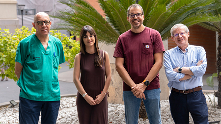
The research was led by Dr Irene del Canto Serrano, from the Experimental Cardiac Electrophysiology Research Group at INCLIVA and Associate Professor in the Department of Electronic Engineering at the University of Valencia, and by David Moratal, Professor in the Department of Electronic Engineering at UPV. Also involved were Dr Francisco Javier Chorro Gascó and Dr Vicente Bodí Peris, coordinators of the Experimental Cardiac Electrophisiology Research Group and the Translational Research Group in Ischaemic Heart Disease and INCLIVA, respectively. Both are cardiologists at the Hospital Clínico Universitario de València and full professors at the Faculty of Medicine of the University of Valencia. All researchers are members of CIBERCV (the Biomedical Research Networking Centre for Cardiovascular Diseases) of the Instituto de Salud Carlos III. Dr María Pilar López-Lereu and Dr José Vicente Monmeneu, from the Cardiac Imaging Unit of Ascires Biomédico Group, also contributed to the study.
Previous study in a healthy population and use of advanced imaging techniques
The starting point for this research was a previous study conducted in a healthy population, which established normalised reference values for myocardial strain parameters obtained by cardiac magnetic resonance imaging. "Having these normal values available made it possible, in the present study, to accurately compare the results obtained in patients with chronic infarction and detect subtle deviations that would not be apparent using conventional techniques. This approach allows for a more precise and quantitative assessment of cardiac function, overcoming the limitations of traditional measures such as ejection fraction," notes Professor David Moratal.
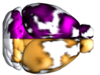
"This study has analysed how cardiac dysfunction can be detected in regions of the heart distant from the area directly affected by a previous infarction, using advanced imaging techniques," says Dr Irene del Canto. Specifically, the researchers used cardiac magnetic resonance imaging and a tool called feature tracking, which enables precise quantification of how the heart muscle deforms during the cardiac cycle. This methodology provides a more sensitive assessment of cardiac function than conventional methods. The research was conducted in patients with chronic myocardial infarction, analysing cardiac MRI images to identify functional alterations that are not apparent in regions of the heart that appear otherwise healthy.
The study was funded by the Conselleria d'Educació, Investigació, Cultura i Esport (CIGE/2023/88), the Valencian Innovation Agency (Generalitat Valenciana) (INNCAD00/19/085), and the Instituto de Salud Carlos III/European Regional Development Fund (CB16/11/00486, PIE15/00013).
Article reference: Del Canto, I., Lopez-Lereu, M. P., Monmeneu, J. V., Chorro, F. J., Bodí, V., & Moratal, D. (2026). Assessment of remote ventricular dysfunction by cardiac magnetic resonance feature tracking strain in chronic infarction. Clinical radiology, 93, 107205. https://doi.org/10.1016/j.crad.2025.107205

La mejor actitud que podemos adoptar es la de trat...

The research team observed changes in head circumf...

AtCDF3 gene induced greater production of sugars a...

En nuestro post hablamos sobre este interesante tipo de célula del...

A study conducted by the Sant Pau Research Institute (IR Sant Pau) and...