Single-vesicle imaging and analysis methodologies focus attention towards exosomes research. Recent advances in the single-vesicle analysis have facilitated their application in medical diagnosis and therapies.
The Exosomes Laboratory and Metabolomics platform, led by Dr. Juan Manuel Falcón-Pérez, reported in Bordanaba-Florit et al., Nature Protocols the current and prospective single-vesicle methodologies and also the recent scientific advances achieved thanks to these technologies (Full-text access to a view-only version: https://rdcu.be/cmEWB)
Short abstract:
Since the description of minute bodies found in a piece of cork by Robert Hooke in 1665, both our scientific knowledge and technical abilities have increased enormously. As techniques have become more accurate and intricate, so has our understanding of biological processes and structures. Technological advances in the field of imaging have resulted in the identification of cell structures, such as the mitochondria and nuclei, and the discovery of different levels of cellular complexity. In 1967, Peter Wolf visualised platelet dust in fresh platelet-free blood plasma using an electron microscope; thus, a mammalian vesicle-like structure was described for the first time. Gradually, these vesicles were characterised in more detail.
Nowadays, these secreted vesicles are extensively reported and widely known as extracellular vesicles (EVs). EVs are heterogeneous, nano- to micrometre-sized, bilayer lipid containers secreted by most cell types. They are multi-purpose carriers that can contain a wide variety of cargos such as lipids, proteins, metabolites, sugars, RNA (mRNA, miRNA, siRNA) and even DNA. In mammals, EVs have been found in body fluids like plasma, urine, saliva, breast milk and seminal fluid, among others.
EVs play a key role in pathophysiological processes, however, the heterogeneity of their structure and composition hinders their application in medical diagnosis and therapies. This diversity makes it difficult to establish their exact physiological roles, and the functions and composition of different EV (sub)populations. Ensemble averaging approaches currently employed for EV characterisation, such as Western blotting or “omics” technologies, tend to obscure rather than reveal these heterogeneities. We describe the contributions of these recent technological advances to the characterisation and phenotyping of EVs, examination of the role of EVs in cell–to–cell communication pathways and the identification and validation of EVs as disease biomarkers. Finally, we discuss the potential of innovative single-vesicle imaging and analysis methodologies using microfluidic devices, which promise to deliver rapid and effective basic and practical applications for minimally invasive prognosis systems.
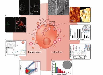
Picture caption:
Schematic overview of the main single-vesicle analysis techniques discussed in this review. Data visualization and single-vesicle interpretation are depicted.
Original publication:
Bordanaba-Florit, G., Royo, F., Kruglik, S.G. et al. Using single-vesicle technologies to unravel the heterogeneity of extracellular vesicles. Nat Protoc (2021).
DOI: https://doi.org/10.1038/s41596-021-00551-z

La mejor actitud que podemos adoptar es la de trat...

The research team observed changes in head circumf...

AtCDF3 gene induced greater production of sugars a...

En nuestro post hablamos sobre este interesante tipo de célula del...

New study finds an association between higher temperatures early in li...