A stroke can damage the brain in many different ways. The traditional view on how a stroke affects brain function exactly is to relate the loss of function, such as motor control, speech, or memory, with the location of the lesion. However, stroke patients also display symptoms that are not easily localized such as chronic pain or depression. How can these, so called, non-specific deficits be understood?
Researchers J.B.G. van Wijngaarden, R. Zucca and PFMJ Verschure of the Synthetic Perceptive, Emotive and Cognitive System (SPECS) research group, in collaboration with professor Simon Finnigan of the University of Queensland in Australia, have tried to answer these important questions that are relevant both to our fundamental understanding of the brain and to advance clinical interventions. They have demonstrated that cortical trauma disrupts distributed neural networks, comprising multiple regions spread across the brain.
These findings are the result of a decade-long focused research program by SPECS, led by ICREA professor Paul Verschure, that uses brain theory and clinical research to target the behavioural and neural consequences of brain deficits such as stroke and to advance science-based neurorehabilitation interventions.
The brain is a complex network of many inter-connected regions that together provide a substrate for perception, cognition, action, and consciousness. To do so, single units of the brain called neurons are constantly communicating with both their local neighbours and distant cells. In [stroke] however, blood supply to the brain is (temporarily) blocked, leading to neuronal cell death. As a result, patients often suffer symptoms directly related to the loss of this brain tissue, including motor deficits (muscle dysfunction) and language disorders (aphasia). But how does stroke affect the crucial communication across the many networks of the brain? And how could possible network disturbances relate to some of the more intractable symptoms of stroke such as pain and depression?
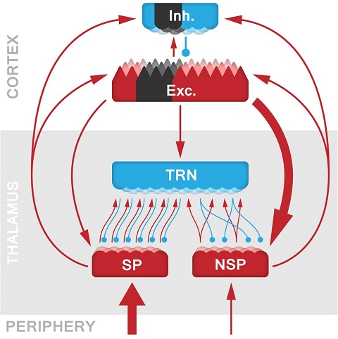
The authors focused on two core brain systems: the [neocortex] and the [thalamus]. The thalamus is the main information gateway for the neocortex and these two structures are bidirectionally coupled via a multitude of direct and indirect pathways. In a previous theoretical study, Verschure and his co-workers had already identified the mechanism behind deficits that occur in this system due to Parkinson’s Disease (PD). In this case it was shown that the characteristic slowing of the EEG that correlates with PD tremor could be traced back to a pathological change in the thalamus, which in turn entrains the neocortex. These disruptions are called Thalamo-cortical Dysrhythmia (TCD).
In this study the question was whether lesions to the neocortex could induce comparable deficits and so account for the intractable non-specific symptoms of chronic pain and depression. This study analysed brain activity data obtained with the electroencephalogram (EEG) of stroke patients within a few days after stroke onset. They looked for specific neural activity signatures within these recordings and compared them with the EEG of other patient groups who suffered from Parkinson’s disease or neurogenic pain (NP) syndrome. They observed that the stroke patient’s EEG showed a remarkable resemblance with those of PD and NP, indicating that thalamic activity was strongly affected by lesions in the cortex.
In order to better understand the pathological change in the interaction between cortex and thalamus, the authors developed a detailed computational model of this system (Fig. 1). Using this model, they were able to specifically identify local cellular, circuit, and network properties that contribute to the development of stroke-induced TCD. Together, these results advance our understanding of the adverse impact of stroke on the function of brain networks. It also sheds light on the indirect, less-understood symptoms of stroke, such as hemispatial neglect or post-stroke pain, suggesting potential new avenues of treatment for stroke and related neurological conditions. Finally, these new neural signatures of stroke can help inform future brain monitoring and improve post-stroke diagnostics in the emerging perspective of network medicine.
Acknowledgments:
This work was supported by the European Research Council under the European Union's Seventh Framework Programme FP7/2007-2013/ERC grant agreement n. 341196 [CDAC], and the Research and Technological Development Programme FP7-ICT-612139 [WYSIWYD] to Paul Verschure.
Reference:
van Wijngaarden JBG, Zucca R, Finnigan S, & Verschure PFMJ (2016), "The Impact of Cortical Lesions on Thalamo-Cortical Network Dynamics After Acute Ischaemic Stroke: A Combined Experimental and Theoretical Study", PLoS Computational Biology 12(8): e1005048.

La mejor actitud que podemos adoptar es la de trat...

The research team observed changes in head circumf...

AtCDF3 gene induced greater production of sugars a...

En nuestro post hablamos sobre este interesante tipo de célula del...
The finding is the first direct observation of how the most abundant p...