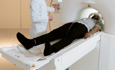
Magnetic resonance imaging (MRI) is an essential imaging modality when evaluating musculoskeletal neoplasms - abnormal tissue masses. Due to its high soft tissue contrast and its ability to acquire multimodal images, MR images provide different information. On the other hand, positron emission tomography (PET) is a fundamental tool in the diagnosis of various malignant neoplasms and offers quantitative molecular and physiological information. The use of both modalities in combined PET / MRI systems makes them especially suitable for evaluating skeletal metastases.
Experts in nuclear medicine and radiology, some of them belonging to 13 of the most important PET / MRI centers worldwide, have shared the experience gained in the use of these imaging modalities to evaluate skeletal metastases. As a result, they have made a statement of opinion on the usefulness of the combination of both modalities when obtaining images of skeletal metastases, thus establishing that “PET / MRI with certain radiotracers (contrasts used in PET) can provide advantages key on the results offered by the PET system combined with computed tomography in the evaluation of bone metastases in several primary neoplasms ”.
URJC professor Ángel Torrado-Carvajal has participated in this study, published in the scientific journal European Journal of Nuclear Medicine and Molecular Imaging and endorsed by various scientific societies in different countries. The researcher notes that, for the evidence in this statement, “we searched the peer-reviewed literature related to the use of PET / MRI in the context of bone metastases. In addition, the expert opinions, practices and protocols of the main research institutions conducting research on PET / MRI of skeletal metastases were considered ”.
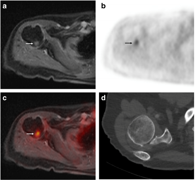
The group of experts concludes in its statement that PET / MRI is positioned, therefore, as the advantageous option for the early and accurate diagnosis of this type of cancer. "This system allows us to observe, in a non-invasive way, both the shape of an organ and its function, as well as the extension of cancer cells, a fact that makes it the best 'tracker' of incipient metastasis", explains Torrado-Carvajal, who emphasizes that "this technology integrates, in a single hybrid device, the two most powerful diagnostic tools at present to detect tumor spread: Positron Emission Tomography (PET) and high-field Magnetic Resonance (MRI)".
A unique system for early detection and customization of treatments
PET / MRI represents a unique model of diagnostic imaging. It combines, in a single exploration, the diagnostic information of the image of the biochemical and molecular processes offered by Positron Emission Tomography (PET) with the morphological and morphofunctional characterization of the tissue offered by Magnetic Resonance (MRI). Therefore, the experts assure that “it should be considered for the staging of neoplasms where there is a high probability of metastatic bone disease based on the characteristics of the primary neoplasm, high clinical suspicion and in the event that the presence of bone metastases has an impact in the management of the patient ”. Thanks to this technology,
In this sense, the PET / MRI system is positioned as the current technology capable of providing greater diagnostic security and more guarantee of success in the treatment of this type of cancer. A technology that opens the possibility of applying a personalized treatment directed towards specific lesions and maintaining better control of the evolution of the treatment, all with less radiation.
This work has been funded by several research projects, among which is the Young Researchers Project directed by Professor Ángel Torrado-Carvajal (Ref. M2166, MIMC3-PET / MR), funded by the Community of Madrid and the Rey University Juan Carlos.

La mejor actitud que podemos adoptar es la de trat...

The research team observed changes in head circumf...

AtCDF3 gene induced greater production of sugars a...

En nuestro post hablamos sobre este interesante tipo de célula del...

New study finds an association between higher temperatures early in li...