Researchers from ICFO and IDIBAPS go one step forward in the understanding of the molecular mechanisms involved in the appearance and development of autoimmune diseases of neuronal cells, such as anti-NMDAR encephalitis. Thanks to a type of microscopy called STORM, they have revealed how the antibodies of patients can modify the function of NMDA receptors, altered in this disease. In the study, published in Cell Reports, ICFO researchers Laurent Ladépêche, Shreyasi Thakur, Irina Suárez, Joseph Steven Borbely, Ángel Sandoval, Lara Laparra, led by Prof. Melike Lakadamyali, in collaboration with IDIBAPS researchers Jesús Planagumà, Makoto Hara, directed by Prof. Josep Dalmau, have participated.
The N-methyl-D-aspartate receptor (NMDAR) is a glutamate receptor, made up of two types of subunits (GluN1 and GluN2), found in nerve cells. This type of receptor plays a key role in neuronal synaptic plasticity and memory function and has been linked to various pathological conditions (e.g. schizophrenia, Parkinson’s disease).
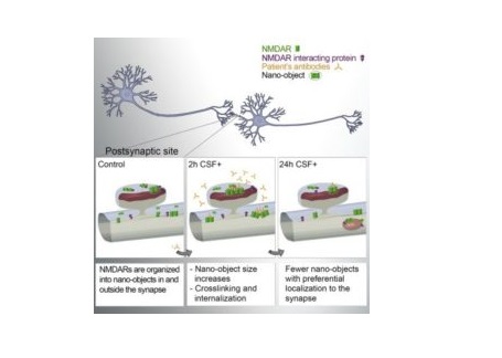
Now, the anti-NMDAR encephalitis is a recently identified severe autoimmune neuropsychiatric disorder characterized by changes in behaviour, psychosis, decrease of memory, seizures, stereotyped movements, autonomic instability, and coma. This disorder is caused by patients’ antibodies, which induce the cross-linking and internalization of NMDA receptors inside the cell, thus altering its function. However, the synaptic events leading to this depletion/internalization of NMDAR are poorly understood.
In a recent study published in Cell Reports, ICFO researchers Laurent Ladépêche, Shreyasi Thakur, Irina Suárez, Joseph Steven Borbely, Angel Sandoval, Lara Laparra, led by Prof. Melike Lakadamyali, in collaboration with IDIBAPS researchers Jesús Planagumà, Makoto Hara, led by Prof. Josep Dalmau, have reported on the nature and timeframe of the processes that take place at the synapse prior to the depletion of NMDA receptors of the cell surface..
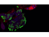
In their study, the team of scientists used single-molecule localization microscopy, in particular stochastic optical reconstruction microscopy (STORM), to reveal how the NMDA receptors distribute along the neuronal dendrites. That is, they were able to visualize the nanoscale organization of the receptors and quantify its relative changes under pathological conditions. They showed that, under normal conditions, NMDARs form nano-sized clusters, which are significantly bigger in the synapses. In presence of the patients’ NMDAR antibodies, the receptors tend to aggregate in those nano-objects, increasing in size and receptor content, both inside and outside the synapses. They also observed that this clustering of receptors, which leads to their internalization and depletion from the cell surface, is specifically affecting one of the subtypes of NMDAR (containing the GluN2B subunit). In addition, they could demonstrate that activating a partner protein of the NMDA receptor, the Ephrin B2 receptor, could partially compensate the deleterious effects of the patients’ antibodies.
To better understand the mechanisms underlying those observations, the scientists carried out computer simulations to model the behaviour of these receptors along the neuronal dendrites in presence of the patients’ antibodies. They could effectively recapitulate their experimental results, allowing them to extract the key parameters leading to the antibody-induced alterations. They revealed that the disruption of protein-protein interactions, such as the one existing between NMDA and Ephrin B2 receptors, is playing an important role in the sequence of events leading to the disease pathogenesis.
The results of this study shed new light on the nanoscopic processes by which patients’ antibodies can alter the function of a targeted protein, opening a new window into understanding the molecular mechanisms of autoimmune disorders. Future studies aim to determine if other interacting partners with NMDAR could be disturbed by patients’ antibodies in order to discover new potential treatment targets.

La mejor actitud que podemos adoptar es la de trat...

El equipo de investigadores observó cambios en el...

El gen AtCDF3 promueve una mayor producción de az...

En nuestro post hablamos sobre este interesante tipo de célula del si...
Curapath se complace en reconocer el importante avance logrado por Mac...