Researchers at the Centre for Genomic Regulation (CRG) in Barcelona have contributed to the creation of new guidelines to improve the clarity, interpretability, and reproducibility of microscopy images and their associated data analysis in scientific publications.
A group formed by 58 scientists from 48 institutes address an urgent need for unified standards to enhance the reproducibility of images. Dr. Nadia Halidi, member of the working group responsible and Head of the Advanced Light Microscopy Unit at the Centre for Genomic Regulation, says: “We are addressing an age-old problem. Even the first users of the microscope, Robert Hooke and Antoni van Leeuwenhoek, didn’t disclose full methodological details. As technological improvements in the field gain pace, it’s more important now than ever to create standardised guidelines and avoid introducing unintentional errors and delays in scientific publications.”
The international microscopy community addressed this challenge by working together for the last two years and creating the first unified guidelines of its kind. Their checklist, published today in a paper the journal Nature Methods, has been designed to help any researcher prepare and analyse light microscopy data for publications.
The checklists address key recommendations for image formatting, annotation, colour selection, data availability, and reporting image analysis workflows. If used accordingly, the authors believe science will benefit from microscopy data that are clearer, standardized, and more reproducible.
Dr. Halidi recalls first thinking of the importance for new guidelines during a personal experience during her doctoral studies: “While attempting to replicate a former student’s work, I grappled with significant hurdles due to the lack of adequate documentation and clarity in image acquisition and data analysis," she says. "The absence of comprehensive documentation often forces us to start from scratch, wasting precious time and resources, and may also compromise reproducibility of the data obtained previously. These checklists are an important response to that and will benefit researchers around the world.
Dr. Halidi is confident that the guidelines will be adopted and serve the scientific community: "Change begins with awareness. We need to disseminate this information not just among PhD students but across all research levels." As for the ultimate goal of universal research reproducibility, Dr. Halidi adds: “Hopefully we will get there. It's a collaborative effort involving journals, institutions, core units and individual scientists.
The creators of the guidelines recognise that, as technology and research progress, the proposed checklists will require regular updates. The aim is to have the guidelines continuously refined by the microscopy community and adapt to future challenges to keep up pace with technological innovation.
The guidelines were developed by the Image Visualization and Analysis working group of QUAREP-LiMi, an international working group of light microscopy imaging experts from academia and industry. The current guidelines were drafted by experts from forty eight organisations, led by Helena Jambor at TU Dresden and Christopher Schmeid at Human Technopole. The working group includes members of academic research institutions such as NASA Ames Research Center or Karolinska Institute as well as industry leaders such as Nikon Instruments and Carl Zeiss and more. Dr. Halidi was the leading contributor to this work in Spain.
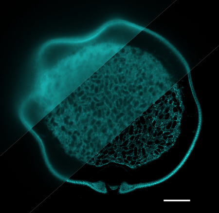
Image: A pollen grain captured with 3 different imaging modalities. From top left to bottom right: widefield, confocal and airyscan microscopy techniques (scale bar: 10 µm). Credit: Nadia Halidi/Centro de Regulación Genómica

La mejor actitud que podemos adoptar es la de trat...

The research team observed changes in head circumf...

AtCDF3 gene induced greater production of sugars a...

En nuestro post hablamos sobre este interesante tipo de célula del...
Many repetitive regions of the genome have been considered “junk DNA...