The main areas of research at the bench focus on finding biomarkers for early detection of the disease, on understanding the molecular mechanisms of the tumor, and on strategies for combatting the disease, such as virotherapy and drugs targeted at the cells that surround the tumor.
Pancreatic cancer is one of the most aggressive tumors of the digestive system and with one of the worst prognoses. Only 5% to 10% of patients survive 5 years after diagnosis. Patients often do not present symptoms until the disease is at an advanced stage and affects other organs, which delays detection. This, together with the lack of an efficacious therapy, explains the poor outcome in this type of tumor.
“Although the term pancreatic cancer includes different tumors, we are normally talking about pancreatic ductal adenocarcinoma, which accounts for 90% of cases”, explains Cristina Fillat, head of the IDIBAPS group Gene therapy and cancer.
All over the world, researchers are working to find new treatments for this cancer and ways of preventing it, as well as identifying the mechanisms that prevent treatments from being successful. Notable areas of research focus on early detection by studying genomic and epigenomic abnormalities that can be used as biomarkers, and on treatment strategies such as improving existing chemotherapy drugs, stimulating the immune system, and immunotherapy to fight the tumor, and advanced therapies.
New biomarkers to improve diagnosis and prognosis
Early detection of pancreatic cancer increases patient survival. “Diagnosis is currently made using invasive techniques such as biopsy or ultrasound-endoscopy-guided fine-needle aspiration”, says Meritxell Gironella, a researcher from the IDIBAPS group Gastrointestinal and pancreatic oncology, directed by Antoni Castells. “We, however, look for biomarkers to diagnose and follow the evolution of the cancer or predict its response to treatment in a noninvasive manner, using a blood sample, i.e., a liquid biopsy”.
Specifically, Gironella and her team study small molecules of non-coding RNA known as micro-RNA. First, they analyze the expression of these molecules in samples of surgical tumor tissue or biopsies from patients. They then select the micro-RNA that presents a greatest abnormality and validate the results in cells obtained via fine-needle aspiration of the pancreas and in plasma samples. “In comparison to tumor tissue, we find little genetic material from the tumor cells in the plasma”. The lack of material makes it harder to detect abnormalities. This means that we need to find micro-RNA with sufficiently pronounced changes to be detected in this type of sample”, explains the researcher.
Another area that the scientists are working on is the study of intraductal papillary mucinous neoplasm, cystic lesions of the pancreas that can become malignant. “The question is not only to diagnose these lesions as early as possible but also to predict which ones will progress to cancer and which will not. The differences in the micro-RNA expression profile can help us to classify them”, says Gironella.
Understanding the disease to cure it
Molecular abnormalities, however, also provide information on what is happening inside the tumor. “In in-vitro models, we compare how the cells behave when we inhibit or suppress some of these micro-RNAs, and we study them based on which mechanisms do this. This allows us to identify the role they play in the development of this cancer and to determine which of them could be used as therapeutic targets”, says Gironella. Micro-RNA, however, may also help to potentiate the effects of chemotherapy. “Tumors have the ability to adapt and develop resistance to treatments. This is why we evaluate the efficacy of combining conventional therapy with the inhibition of certain micro-RNAs”.
Fillat and her team focus their research on a protein involved in the processes of transforming normal cells into cancer cells. “Some years ago, as part of a study of Down syndrome, we began to study the protein DYRK1A, coded for by a gene located on chromosome 21”, explains the researcher. “The results suggested that this protein played a role in the cellular signalling and proliferation processes that are often altered in cancer cells. This led us to analyze the expression and functionality of DYRK1A in pancreatic tumors. We also observed that inhibiting this protein might contribute to controlling the progression of the cancer”.
The tumor environment is also important
Nevertheless, expression of DYRK1A is not just limited to cancer; the protein is also expressed in the surrounding cells, specifically the fibroblasts. “In a pancreatic tumor, we find fibroblastssome neoplastic cells, but we also observe a large number of fibroblasts and other cells in the neighborhood”, explains Fillat. The fibroblasts are not uniform and different populations can be distinguished, which can play opposing roles in controlling tumor proliferation. On the one hand, there is a type of fibroblast, known as iCAF, which is more proinflammatory and which promotes growth of the tumor, and on the other, there is a subpopulation, known as myCAF, which may inhibit progression of the tumor.
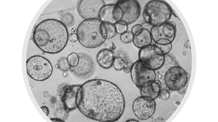
The researchers do not know how DYRK1A fits into this puzzle, but they have developed a preclinical model that allows them to study the interaction between the fibroblasts and the tumor in order to understand it. “We have established lines of organoids, i.e., three-dimensional cultures obtained from patients’ tumor cells, which we culture together with the fibroblasts. This co-culture allows us to better approximate the tumor in the body”, adds Fillat.
Eva Vaquero, a researcher with the IDIBAPS group Gastrointestinal and pancreatic oncology, leads an area of research that focuses on the fibroblasts in the fibroinflammatory tissue, or stroma, surrounding the tumor. These cells may be a therapeutic target for preventing the onset and development of the cancer in susceptible cases with the disease. “Drugs with antifibrogenic activity may modulate the characteristics or phenotype of the fibroblasts. They could thus transform the iCAF proinflammatory fibroblasts to myCAF and, therefore, provide a potential way to suppress their protumoral properties, i.e., the properties that promote the tumor”.
Vaquero and her team are investigating the effect of these compounds in a mouse model that reproduces the characteristics at the tissue level in human pancreatic adenocarcinoma. “The mice that develop the tumor are carriers of the mutation in the KRas gene in the pancreatic cells and are fed a high-fat diet. Through the diet, the animals are given the compound nintedanib for several weeks; this drug is used to treat pulmonary fibrosis and attenuates the protumoral properties of the fibroblasts and delays development of the cancer”. These results are not transferrable to clinical practice, but they show the therapeutic potential of strategies aimed at the fibroblasts.
Viruses against cancer
Another notable therapeutic strategy is virotherapy. This is based on the use of oncolytic viruses, which cause the rupture, or lysis, of the cancer cells without affecting healthy cells. Not all viruses, however, present a natural affinity for tumor cells. Adenoviruses, which are used by Fillat’s group, are an example of this type of virus, but they need to be genetically modified. “Once this obstacle has been overcome, the adenoviruses are a highly potent therapeutic tool, as once inside the cell, they multiply and generate new therapeutic elements in the tumor itself, while they eliminate it. This means that there is no need for continuous and repeated administration of the treatment”, explains the researcher. “The fibroblasts and the extracellular matrix surrounding the tumor, however, act as a kind of barrier that limits the propagation of the viruses and, as a result, prevent the elimination of all the tumor cells”.
Virotherapy also faces other challenges, such as the route of administration. The main route of interest is systemic. That is, all the routes that allow the treatment to pass into the circulatory system and distribute the treatment throughout the body. “However, most people become exposed to adenoviruses during their lives and, as a result, have antibodies that can neutralize them before they reach the tumor or its metastasis”. To solve this problem, Fillat and her team are seeking to develop polymers that will encase the virus and act as a shield. “Once they reach the target, the camouflage system would disappear and the adenoviruses could then act against the cancer”, she concludes.
It should also be noted that adenoviruses can potentiate the effect of other treatments, such as immunotherapy. After breaking the cell membrane, the proteins of the oncolytic virus and other components are released from the tumor cells and generate immunogenic death. This constitutes an alarm signal and a cry for the dendritic cells of the immune system to act by capturing more tumor cells. The immune cells then migrate to the lymphatic nodes where they activate the cytotoxic T cells, which will return to the site of the cancer, recognize the antigens of the tumor cells and attack them.
Fillat, Gironella, and Vaquero agree in highlighting precision medicine as the future of research into pancreatic cancer. “Precision medicine means studying the tumor at the molecular level before treating it. We need to have a clear idea of which molecules we can act on, which markers may indicate the most effective treatment, and this depends on each patient; we do not have a formula that fits everyone”, says Vaquero.
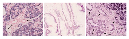
Image: Images of healthy tissue (left), intraductal papillary mucinous tumor (center) and pancreatic ductal adenocarcinoma (right) where miR-181a-5p microRNA expression is observed. Depending on the intensity of expression, the staining is either blue/violet (high) or light pink (low). The arrows show ductal cells and the scale bar is 50 microns.

La mejor actitud que podemos adoptar es la de trat...

The research team observed changes in head circumf...

AtCDF3 gene induced greater production of sugars a...

En nuestro post hablamos sobre este interesante tipo de célula del...

New study finds an association between higher temperatures early in li...