Growing research in photostimulation indicates that exposure to light can have a positive impact on health-related problems such as spring asthenia, circadian rhythm disruption and even bipolar disorders and Alzheimer’s.
However, the extent and location of changes in brain areas caused by the exposure to monochromatic light remain largely unknown. To move forward in this field, a group of researchers from the Terrassa School of Optics and Optometry (FOOT) of the Universitat Politècnica de Catalunya-BarcelonaTech (UPC) has conducted a pilot study. They have demonstrated that just one minute of blue, green or red-light exposure modifies the functional connectivity of a wide range of neural networks or visual and non-visual brain regions.
According to the researchers, each one of the brain connectivity pattern appears to be best arranged to perform better on tasks associated with specific cognitive domains: blue-light exposure activates areas associated with attention and circadian rhythm, green-light exposure improves visual attention and red-light exposure influences areas such as memory.
Recently published in the journal Scientific Reports (Nature), the study has been coordinated by researcher and FOOT professor Marc Argilés, with the collaboration of students Bernat Sunyer and Silvia Arteche, from the UPC’s doctoral programme in Optical Engineering. Funded by the Official Association of Optometrists of Catalonia (COOOC), it has involved the Barcelonaβeta Brain Research Center (BBRC), created by the Pasqual Maragall Foundation, and doctor Cleofé Peña Gómez, from Netdatica. The study has also had the support of the Catalan Association of Optometry and Vision Therapy (ACOTV).
Research development
The pilot study involved seven subjects—four women and three men—aged between 21 and 33 receiving 1-minute exposure to one of three wavelengths (blue, green and red), which was done using light exposure instruments lent by the Centre d’Optometria Mataró. The instruments had been studied and characterised optically (wavelength, irradiance and photon density) at the FOOT, with the collaboration of professor Elizabet Pérez.
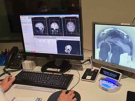
Additionally, functional magnetic resonance measurements were performed on the subjects, which allowed the researchers to observe how brain connectivity underwent short-term changes in all participants and in the same brain regions after just 60 seconds of light stimulation: “We observed a global decrease in functional connectivity (FC) in all the networks but the salience network after blue-light exposure, a global increase in FC after green-light exposure, particularly noticeable in the left hemisphere, and a decrease in FC on attentional networks coupled with an FC increase in the default mode network after red-light exposure”, explain the authors.
“The study opens new avenues for better understanding the impact of light stimulation on brain function and its use to treat not only visual dysfunction, but also depression symptoms, circadian rhythm disruption, migraine and memory or attention disorders”, conclude the researchers.
Image: Images showing the results of the tests that made it possible to observe the changes in brain connectivity.

La mejor actitud que podemos adoptar es la de trat...

The research team observed changes in head circumf...

AtCDF3 gene induced greater production of sugars a...

En nuestro post hablamos sobre este interesante tipo de célula del...

Telum Therapeutics, a biotechnology company leveraging proprietary met...


