In a typical human lifetime, the cells in our bodies will divide countless trillions of times. Each division depends on a molecular machine called the mitotic spindle, cellular scaffolding at the molecular scale which helps separate our genetic material in opposite directions so that each new cell inherits the correct DNA.
Despite its importance, the essential ingredients required to build a stable, functional spindle have been a longstanding mystery. The knowledge could help explain when, where, why and how cell division goes wrong, a phenomenon closely linked to diseases like cancer. It could also help develop new materials developed using inspiration from nature.
A study published today in Proceedings of the National Academy of Sciences shows that just two specific proteins are required. Researchers at the Centre for Genomic Regulation (CRG) in Barcelona and the University of Cambridge found that kinesin-5 and dynein have evolved the perfect properties for the task.
“It’s remarkable how simple it is to form the basic shape of a spindle,” says ICREA Research Professor Thomas Surrey, senior coauthor of the paper and researcher at the Centre for Genomic Regulation. “Our study suggests you only need very few core components. All the other factors in the cell are like add-ons, refining or stabilising a basic system that already works. No extra frills required.”
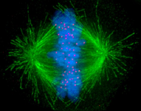
The spindle is made of slender rods known as microtubules. Each of these are only about 25 nanometres wide, roughly 1/4,000th the thickness of a human hair. During division, each human cell builds and tears down the spindle in under an hour, while microtubules themselves are being created and destroyed in minutes. At any given moment in our bodies, millions of cells are performing this feat simultaneously.
‘Crosslinking’ proteins are another key ingredient. These molecules create a physical bridge between two microtubules, making each rod move or stay in specific positions. This helps give shape to the spindle, ensuring the machine is stable and properly aligned to separate chromosomes.
When the spindle has two clear poles – each in opposing directions – chromosomes are separated correctly and two new cells are born. When it has multiple poles, it can cause chromosome segregation errors that cause cell division to fail, which in turn can cause cancer or many other types of disease.
Using computer simulations, the researchers demonstrate that creating a stable, two-pole spindle is simpler than expected. By changing traits like the speed and binding strength of different proteins, the researchers could see what conditions produce stable two-poled structures in human cells. Kinesin-5 and dynein worked together to create the classic spindle shape which can then pull DNA apart.
The findings emerged from an open-source software platform called Cytosim, developed by Dr. Franҫois Nédélec, co-senior author of the study and researcher at the University of Cambridge. The computer model simulates the mechanical and chemical behaviour of microtubules and their associated proteins.
“It’s easier to explore many different protein designs and properties in simulations than in experiments. This allows us to understand, in quantitative terms, why these proteins have evolved to what they look like today,” says Dr. Wei-Xiang Chew, postdoctoral researcher at the CRG and first author of the study.
“Their synergy is critical so that chromosomes can be moved along two opposite directions toward the exact two poles of a healthy spindle, in contrast to multi-pole spindles that are a hallmark of cancer,” he adds.
The study can boost cancer research efforts and the study of other diseases caused by abnormal cell division. The computer models can help explain how to avoid multi-pole spindle formation. For example, it can be used to predict how certain mutations in DNA can change a motor’s speed or disrupt its spindle assembly.
Understanding the spindle’s core design can also help scientists rebuild simplified spindle versions in the lab, something Thomas Surrey’s lab at the CRG is already doing, with the potential to shine light on which drug targets matter most for tackling cancer and other diseases tied to abnormal cell division.
The research can also resonate beyond cell biology. The researchers compare the proteins working in cell division to the components of materials that can power themselves, also known as active matter, a field with industrial applications ranging from the design of nano-robots to self-assembling materials.
With traditional materials, things only move if you push them externally. But in active matter, individual building blocks each use up a little bit of energy on their own to sustain the continual internal motion which lets the system reorganise itself into patterns like spirals or vortices. In this case, the materials use ATP, the building block of life used to generate energy, to form the spindle and help cells divide.
“Active materials are responsive, adaptive and can also have the capacity to repair themselves. Spindles are self-repairing, adaptive, and out of thermodynamic equilibrium, exactly the qualities that fascinate material scientists. Borrowing nature’s trick could one day help build new smart materials that adjust and fix themselves without external input,” concludes Dr. Surrey.
Image: Image of the mitotic spindle in a human cell showing microtubules in green, chromosomes (DNA) in blue, and kinetochores in red.

La mejor actitud que podemos adoptar es la de trat...

The research team observed changes in head circumf...

AtCDF3 gene induced greater production of sugars a...

En nuestro post hablamos sobre este interesante tipo de célula del...

Telum Therapeutics, a biotechnology company leveraging proprietary met...


