The Parc Taulí started this July one multicenter clinical trial for the validation of a artificial intelligence software that detects lung cancer in unreported chest x-rays. This tool, named Optimal XR, aims to prevent potentially malignant lung nodules from continuing to go unnoticed that are currently not diagnosed in time because the x-rays have not been examined by a radiologist.
The study, led by the pulmonologist and researcher emeritus of the Parc Taulí Research and Innovation Institute (I3PT) Eduard Monsó, aims to confirm the effectiveness and safety of the algorithm, as well as validate its use in a real hospital setting and generate the evidence to be able to apply for approval to commercialize it.
The clinical trial will have an estimated duration of sixteen months—ten recruitment and six clinical follow-up—with additional passive follow-up three years after its completion. During the course, nearly 3.000 chest x-rays will be analyzed in real time from the database of the Digital Medical Imaging Center (CIMD) of Parc Taulí that are not necessarily related to respiratory diseases, but may have been taken by other medical reasons. "The study will consist of using the algorithm to identify pulmonary nodules in these radiographs, but not all of them will contain them. The nodules detected by the system will be reviewed by a radiologist and, if confirmed, will be referred to the Pneumology Service for a more detailed evaluation and follow-up," he explains. monsoon.
Optimal XR and lung cancer
Lung cancer is the leading cause of cancer death worldwide. In Catalonia alone, in 2023, almost 5.000 new cases and more than 3.400 deaths were recorded due to this disease. The late appearance of its symptoms, however, means that 80% of lung cancers go unnoticed and are diagnosed in too advanced stages, when the survival rate after five years is already less than 20%. Timely detection not only significantly increases the survival rate — up to 60% — but also improves the living conditions of people who suffer from it.
Currently, early detection of lung cancer is a major challenge for Primary Care, where thousands of chest radiographs are performed each year on a significant proportion of patients as part of standard evaluations or as diagnostic procedures that need not be related to an initial suspicion of this disease. However, due to the growing lack of specialized radiologists, it is not possible to examine these X-rays with the efficiency that would be desired, with the possible risk of failing to identify cancerous lung nodules in the early stages that are later detected when the disease is already in advanced stages.
"We saw the need to develop and implement a solution that could accurately and massively analyze these chest X-rays coming from Primary Care, thereby detecting high-risk cases and directing them into the radiology workflow to dramatically improve identification and intervention early", says Monsó.
In this context, from the Parc Taulí University Hospital i theHospital Universitari Vall d'Hebron, together with the technology center Eurecat, was developed Optimal Lung, a clinical decision support system based on artificial intelligence that integrates two algorithms for the detection of pulmonary nodules: Optimal XR, which focuses on radiographs from Primary Care and emergency departments, to avoid missing cancerous lung nodules; i Optimal CT, which focuses on CT scans to cover all levels of lung cancer diagnosis.
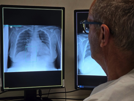
Optimal XR is an artificial intelligence software that uses deep learning technology — in English deep learning — to analyze radiographs. The algorithm processes all x-rays taken at the hospital center and identifies those that have a high probability of containing lung nodules, based on more than a thousand real x-rays it has previously trained on. Detected radiographs are sent directly to a radiologist to review and confirm the presence of nodules. In this way, the work of radiologists is made easier and the early and timely detection of possible lung problems that until today could have been unnoticed is improved.
In the development of this solution, departments from different clinical specialties such as radiology, pneumology and oncology have participated, as well as engineers, experts in information systems and innovation staff. The ultimate goal will be to implement Optimal XR in other hospitals, with a special focus on Primary Care settings where it is now most needed. Later, according to Monsó, "the intention of the team is to incorporate it in other countries where they do not have the attention or reading of chest radiographs as developed as we have here".

La mejor actitud que podemos adoptar es la de trat...

The research team observed changes in head circumf...

AtCDF3 gene induced greater production of sugars a...

En nuestro post hablamos sobre este interesante tipo de célula del...

Telum Therapeutics, a biotechnology company leveraging proprietary met...


