Oocytes are immature egg cells that develop in almost all female mammals before birth. The propagation of future generations depends on this finite reserve of cells surviving for many years without incurring damage. In mice, this can be a period of up to eighteen months, while in humans it can last almost half a century, the average time between birth and menopause. How the cells accomplish this remarkable feat of longevity has been a longstanding question.
Researchers at the Centre for Genomic Regulation (CRG) in Barcelona have discovered a new mechanism which explains how oocytes remain in pristine conditions for decades without succumbing to the wear and tear that would cause other cell types to fail. The findings, reported today in the journal Cell, represent a new frontier to explore unexplained causes of infertility.
The researchers looked at protein aggregates, which are clumps of misfolded or damaged proteins. If left unchecked, these harmful substances accumulate in the cytoplasm and have highly toxic effects. Protein aggregates are known to accumulate in neurons and their effects have been linked to several neurodegenerative diseases. Cells usually manage aggregates by breaking them down with specialized enzymes. They can also divide into two new cells, concentrating aggregates in one of the cells and sparing the other.
But oocytes are not like the other cells. Their long life means they cannot dissipate toxic substances through cell division. Constantly breaking down protein aggregates is an inviable strategy, as it requires using a high amount of energy that may not be available. Oocytes also have the job of donating their entire cytoplasm to an embryo after fusing with a sperm, and so prefer to reduce their metabolic activity, a strategy which avoids generating by-products which can damage the maternal DNA and compromise future reproductive success. This makes oocytes particularly sensitive to the effects of misfolded or damaged proteins.
However, “in contrast to the tens of thousands of papers on protein aggregation in neurons, how mammalian oocytes cope with protein aggregation is essentially unstudied, despite having the same problem of being long-lived and non-dividing”, explains Dr. Elvan Böke, Group Leader of the Oocyte Biology & Cellular Dormancy programme at the Centre for Genomic Regulation and author of the study. “We wanted to explore how oocytes deal with these misfolded or damaged proteins”, adds Dr. Böke.
Patrolling ‘clean-up crews’
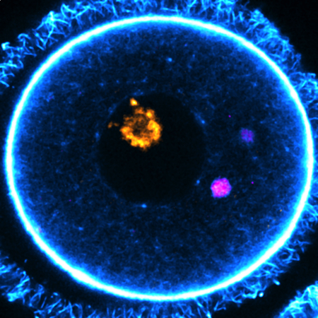
Dr. Böke’s team, led by Dr. Gabriele Zaffagnini, started by collecting thousands of immature oocytes, mature eggs, and early embryos from adult mice. Using special dyes, they observed how the protein aggregates behave in real-time using a technique called live-cell imaging. They also used electron microscopy to get a closer look and see nanoscopic details inside cells, work that took five and a half years to complete.
The researchers discovered special structures in the oocytes which they named EndoLysosomal Vesicular Assemblies – or ELVAs for short. These structures – there are about 50 per each oocyte - roam the cytoplasm, where they capture and hold onto protein aggregates, rendering them harmless. Cells have many subcellular structures known as organelles, which perform jobs much like an organ does in the body. The researchers conceptualise ELVAs as a “superorganelle” because it is a network of many different types of cellular components working together as a single unit.
The study revealed a crucial moment during the oocyte maturation stage, which is when an oocyte converts into a mature egg, preparing for ovulation and possible fertilisation. During this stage, the researchers observed ELVAs moving towards the cell's surface and breaking down the protein aggregates, essentially deep-cleaning the cytoplasm. This is the first observation of the unique strategy oocytes employ to get rid of protein aggregates.
“An oocyte must donate all its cytoplasm to the embryo at fertilisation, so it cannot afford for garbage to accumulate, which would pose an existential risk for its function. In that sense, ELVAs are like a sophisticated waste disposal network or clean-up crew, patrolling the cytoplasm to ensure no aggregates are freely floating. ELVAs keep these aggregates in a confined environment until the oocyte is ready to dispose of them in one fell swoop. It’s an effective and energy-efficient strategy,” says Dr. Zaffagnini, postdoctoral researcher at the Centre for Genomic Regulation.
Protein aggregates may contribute to infertility
Fertility declines with age, and poor oocyte quality is the major cause of female infertility. Global infertility rates are also on the rise, with delayed motherhood being one of the contributing factors. Understanding how oocytes remain healthy, and what causes these strategies to fail with age, is critical for understanding unexplained causes of infertility and open up new avenues for treatment.
The findings of the study suggest that the presence of protein aggregates could interfere with both egg and embryo quality. When the researchers experimentally prevented the ability of ELVAs to degrade protein aggregates during the oocyte maturation process, it led to the formation of defective eggs. When the researchers intervened and “forced” the embryos to inherit aggregated proteins, 3 in 5 (60%) failed to complete very early stages of development.
“A lot of studies have historically focused on one small aspect of why oocyte quality declines, which are meiosis and euploidy. However, a recent review of eleven thousand embryo transfers has shown that the decline in female fertility with age are heavily influenced by other, yet unknown factors. Our research opens a fascinating future direction to explore whether protein degradation, and problems with how they are regulated in oocytes, could explain the age-related decline in embryo health,” concludes Dr. Böke.
Another type of long-lived cell which do not divide yet have to deal with protein aggregates are neurons. The accumulation of the harmful substances in these cells is linked to the development of several types of neurodegenerative diseases including Alzheimer’s. Could ELVA-like compartments also exist in neurons and other cell types? The study opens the door for future research avenues beyond the field of reproduction.

La mejor actitud que podemos adoptar es la de trat...

The research team observed changes in head circumf...

AtCDF3 gene induced greater production of sugars a...

En nuestro post hablamos sobre este interesante tipo de célula del...

Telum Therapeutics, a biotechnology company leveraging proprietary met...


