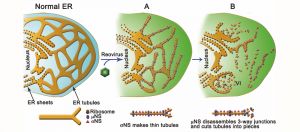
Reoviruses build new organelles (viral factories) in the cytoplasm of infected cells to be able to replicate themselves. This study reveals that the human reovirus modifies and uses endoplasmic reticulum (ER) membranes to produce its replication organelles. The reovirus proteins σNS and μNS are in charge of ER remodeling and the use of this modified ER as scaffold for the assembly of viral factories.
The Reoviridae family causes respiratory and digestive sickness, mainly in children, and has been related to celiac disease in adults. Growing interest in reovirus research is fueled by the observation that this virus can selectively infect and kill cancer cells, which has prompted its use a in cancer therapy. For these reasons, studies of the Reoviridae family have become highly relevant to many aspects of human health.
We know that reoviruses build their viral factories with resources of the infected cell, but the questions which membranes the virus uses for this purpose and how they are modified remained unresolved. The present study resolves this enigma by showing how reovirus remodels ER membranes and how it uses them to build viral factories in very early stages of viral infection.
The reovirus protein σNS is in charge of stretching ER tubules and μNS cuts them into small pieces. These modified ER fragments now serve as scaffold for the assembly of viral factories and supportive structure for the viral replication complex and assembly of new viral particles.
Advanced optical microscopy techniques made it possible to observe how viral proteins remodel the ER structure. Electron microscopy approaches, such as 3D tomography and gold immunolabeling, uncovered a new process of ER remodeling induced by reovirus infection and revealed the importance of these modified ER membranes in the process in the viral replication.
Raquel Tenorio, researcher at the CNB-CSIC and first author of this study, points out that “knowing how and when reoviruses use cellular resources to build their viral factories is a key point in our effort to identify new antiviral targets or cancer therapies”.
This work is the result of a collaboration between the Cell Structure Laboratory at CNB-CSIC, headed by Dr. Cristina Risco, and the group of Dr. Terence Dermody at the Department of Pediatrics at the University of Pittsburgh, USA.
Photo: The reovirus proteins σNS and μNS are in charge of ER remodeling and the use of this modified ER as scaffold for the assembly of viral factories Raquel Tenorio et al., mBio 9:e01253-18 (2018)

La mejor actitud que podemos adoptar es la de trat...

The research team observed changes in head circumf...

AtCDF3 gene induced greater production of sugars a...

En nuestro post hablamos sobre este interesante tipo de célula del...
Acute myeloid leukemia (AML) does more than infiltrate the lungs: it a...