Scientists at the Centro Nacional de Investigaciones Cardiovasculares Carlos III (CNIC) have demonstrated the fundamental role of the hypoxia response in the correct formation of the heart ventricles. Hypoxia, a drop in the levels of oxygen, triggers a complex adaptive response to reestablish the tissue supply of nutrients and oxygen. The key elements of this response are the hypoxia-inducible transcription factors, or HIFs, which mediate the activation of a multitude of genes that guarantee a transitory adaptation to the lack of oxygen.
This pathway plays a well-established role in cancer and tumor metastasis, but its participation in physiological processes has been less explored. The CNIC team, led by Dr. Silvia Martin-Puig, has identified discrete metabolic territories within the embryonic myocardium; the study uncovers the molecular mechanisms through which HIF1 establishes this metabolic boundary between different types of cardiomyocytes to regulate the maturation of the contractile muscle and the establishment of the conduction system. The study, published in Developmental Cell, describes the importance of the hypoxia pathway in an essential physiological process, the formation of the heart chambers, establishing the importance of hypoxia outside of disease settings.
The study findings could help to identify mechanisms underlying congenital heart conditions associated with hypoxia or metabolic alterations, such as preeclampsia or gestational diabetes. Moreover, comments Dr. Martin-Puig, these mechanisms “could be of clinical interest in the treatment of disorders affecting the adult heart. In myocardial infarction, the lack of oxygen, or ischemia, induces the activation of HIF1, which could reprogram the metabolism of the mature myocardium toward an embryonic pattern.” These metabolic adaptations linked to HIF1 activation “could also operate in other cardiomyopathies whose development is associated with energetic alterations.”
Model challenged
Until now, heart metabolism was thought to be dominated by glucose consumption at embryonic stages followed by a switch just after birth to the energetically more efficient fatty acid oxidation, to take advantage of the increased oxygen availability. The research by Dr. Martin-Puig’s team now challenges this model, describing a new point of regulation during gestation related to a decrease in the cardiac levels of HIF1.
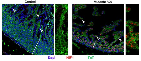
The CNIC research team describe how the distribution of HIF1 in distinct zones of the developing heart establishes compartments with different metabolic programs. “The compact myocardium, which gives rise to the contractile muscle in the mature heart, has high levels of HIF1 and a glycolytic metabolism,” explains first author Iván Menéndez-Montes. In contrast, “the trabecular myocardium, which gives rise to the ventricular conduction system responsible for transmitting cardio-electric stimuli, expresses negligible levels of HIF1 and glycolytic enzymes, instead having a higher mitochondrial activity than the compact myocardium.”
The identified mechanisms confirm the physiological role of HIF1 signaling during heart development, contrasting with its pathological involvement in cancer and pulmonary hypertension
Metabolic program
The authors describe the mechanism through which this metabolic boundary disappears at midgestation (day 14.5 in the mouse), with a sharp drop in the expression of glycolytic enzymes and a simultaneous increase in the number of mitochondria and the activity of genes involved in fatty acid metabolism. This change in the metabolic program coincides with the loss of HIF1 expression in the heart.
The study opens the way to new interventions to improve heart function in distinct situations, including myocardial infarction
Although the scientists are continuing their investigation into the mechanisms that regulate HIF1 fluctuations during development, their work with several genetic models has already demonstrated that maintaining HIF1 activation beyond midgestation causes severe structural defects incompatible with life. Dr. Martin-Puig points out that “the sustained presence of HIF1 maintains a metabolic program based on glucose consumption and causes a sharp decline in the number and activity of cardiac mitochondria, impeding the switch to oxidative metabolism. These alterations to the metabolic program and HIF1 expression in the embryo compromise the contractile capacity of the myocardium and significantly impair heart function.”

La mejor actitud que podemos adoptar es la de trat...

The research team observed changes in head circumf...

AtCDF3 gene induced greater production of sugars a...

En nuestro post hablamos sobre este interesante tipo de célula del...

Telum Therapeutics, a biotechnology company leveraging proprietary met...


