Publishing in the journal Small, the Nanoprobes and Nanoswitches group describe a new way to observe conduction pathways in redox proteins and complexes – in which the transfer of electrons causes a change in oxidation – at the nanoscale.
Electron transfer in proteins is essential in crucial biological processes; cellular respiration, for example, is the oxidation of glucose to CO2 and the reduction of oxygen to water. But even though the basic aspects of electron transfer have been studied thoroughly, so far there has been no tool able to examine the nanoscale electronic pathways by which this transfer happens.
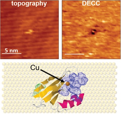
The IBEC group, experts in electrochemical scanning tunneling microscopy, added an alternating current modulation technique to this special kind of microscopy that allowed them to measure the local electronic properties of an electrode, as well as the redox processes occurring at the interface of the electrode and the immersing solution. “This method, Differential Electrochemical Conductance (DECC) imaging, has allowed us to see the reversible oxidation of an iron electrode at the nanoscale,” says first author Montserrat López, a PhD student in ICREA professor Pau Gorostiza’s Nanoprobes and Nanoswitches group. “We’ve also used it to obtain images of a biomolecular redox system, the protein Azurin immobilized on gold.”
In particular, the scientists were able to view submolecular regions with high conductance within the protein that act as pathways for the transfer of electrons. As well as enabling important advances in biochemistry and bionanotechnology, this method will also open new avenues in the design and optimization of molecular electronics, where synthetic compounds are components to build nanoelectronic devices.
—
Source article: M. López-Martínez, J. M. Artés, V. Sarasso, M. Carminati, I. Díez-Pérez, F. Sanz, & P. Gorostiza (2017). Differential Electrochemical Conductance Imaging at the Nanoscale. Small, epub ahead of print
Imagen: Top: Simultaneous high-resolution images: the new method, DECC, is on the right. A single azurin protein displays a submolecular spot with high differential conductance surrounded by a lower conductance region. Bottom: Top-view representation of an Az protein on an atomically flat gold surface. The protein is bound to the surface through a pair of cysteine residues (in green); hydrophobic residues (blue) surround the redox center (brown).

La mejor actitud que podemos adoptar es la de trat...

The research team observed changes in head circumf...

AtCDF3 gene induced greater production of sugars a...

En nuestro post hablamos sobre este interesante tipo de célula del...

Telum Therapeutics, a biotechnology company leveraging proprietary met...


