Clínic-IDIBAPS researchers have led a study that adapts a technique used in the diagnosis of prion diseases, called real-time quaking-induced conversion (RT-QuIC), and combines it with other biomarkers in cerebrospinal fluid and magnetic resonance images with the aim of differentiating Parkinson’s disease from atypical parkinsonisms and improving diagnosis.
Degenerative parkinsonisms are characterised by the misfolding and accumulation of proteins that contribute to neuronal death and the development of the disease. Depending on whether the altered protein is α-synuclein or tau, these disorders can be classified as synucleinopathies, such as Parkinson’s or multiple system atrophy, or as tauopathies, such as progressive supranuclear palsy or corticobasal degeneration. “Despite this, the lack of sufficiently sensitive and specific techniques for detecting α-synuclein is an obstacle to the reliable molecular diagnosis of Parkinson’s disease”, explains Yaroslau Compta, a researcher from the IDIBAPS Group on Parkinson disease and other neurodegenerative movement disorders: clinical and experimental research and co-first author of the study in which researchers from the Parkinson's Unit of the Teknon Medical Center, the Hospital Sant Joan Despí Moisès Broggi, the Hospital General de Granollers and the Hospital Comarcal Sant Jaume de Calella have also participated.
RT-QuIC uses the capacity of the misfolded proteins to transmit the abnormal folding to normal proteins, a phenomenon that increases the quantity of proteins altered to reach detectable levels. “The first study that exploited the prion-like behaviour of α-synuclein was published in 2016. Since then, different laboratories around the world, including our own, have confirmed the high sensitivity of this technique for detecting the protein in samples from patients with Parkinson’s disease, and to a lesser extent, in cases of multiple system atrophy”, states Compta. “The novel aspect of our work lies in the combined use of the detection of α-synuclein through RT-QuIC and of long-chain neurofilament, a fibrous protein that forms part of the structure of neurons, with magnetic resonance images to guide the diagnosis. Even though cases of Parkinson’s usually present high levels of α-synuclein and low concentrations of neurofilaments, often, with the values of each biomarker taken separately, there is not enough to obtain an accurate diagnosis. Brain images alone are not sufficient, either”.
The study, published in the journal Parkinsonism and related disorders, analyses samples of cerebrospinal fluid from 112 participants, between healthy controls and people with different types of degenerative parkinsonisms, including Parkinson’s disease, as well as the surfaces of the mesencephalon and the pons of the volunteers. “According to the results, the three measures together enable Parkinson’s disease cases to be discriminated from the rest of the parkinsonisms with great precision. This differentiation is very difficult based only on the clinical symptoms and has important implications with regard to defining the most appropriate therapeutic strategy for each patient, as well as in predicting the evolution that the neurodegenerative disorder will follow. “In fact, it is important to highlight that, in some cases, the profile of biomarkers obtained at the laboratory did not coincide with the prior clinical diagnosis. This led us to re-examine the patients and classify them anew, which has enabled us to improve their management and care”.
The work has received funding from the Instituto de Salud Carlos III, as well as from the Fundació La Marató de TV3.
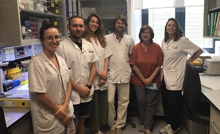
Image: From left to right, Marta Soto, Manel Fernández, Cèlia Painous, Yaroslau Compta, Maria Josep Martí and Ana Cámara, authors of the study.
Reference article
Y. Compta, C. Painous, M. Soto, M. Pulido-Salgado, M. Fernández, A. Camara, V. Sánchez, N. Bargalló, N. Caballol, C. Pont-Sunyer, M. Buongiorno, N. Martin, M. Basora, M. Tio, D.M. Giraldo, A. Pérez-Soriano, I. Zaro, E. Muñoz, M.J. Martí, F. Valldeoriola. Combined CSF α-SYN RT-QuIC, CSF NFL and midbrain-pons planimetry in degenerative parkinsonisms: From bedside to bench, and back again. Parkinsonism & Related Disorders, 2022. 99: 33-41. https://doi.org/10.1016/j.parkreldis.2022.05.006.

La mejor actitud que podemos adoptar es la de trat...

The research team observed changes in head circumf...

AtCDF3 gene induced greater production of sugars a...

En nuestro post hablamos sobre este interesante tipo de célula del...

Telum Therapeutics, a biotechnology company leveraging proprietary met...


