Scientists from the Singular Laboratory of Biofabrication and 3D (bio)printing (BioFabi3D) of the University of Granada (UGR) and ibs.GRANADA have developed a human bioprinted three-dimensional model that recreates the tumor microenvironment of malignant melanoma, the most lethal type of cancer skin.
The research, published in the prestigious journal biofabrication and patented, demonstrates that this model can be used to identify patient resistance to drug treatments and to develop personalized therapies against melanoma.
Conventional cancer models do not accurately reproduce the tumor microenvironment, which limits their usefulness in research and development of new treatments. 3D bioprinting represents an excellent tool to overcome these limitations, since it allows the generation of complex models where all the structures that give rise to tumor tissue are incorporated without the need to use experimental animals. Thanks to this technology, UGR scientists have succeeded in creating a multicellular model of malignant melanoma with the three layers of the skin, which mimics the patient's native tumor niche.
This bioprinted model is comprised of patient-derived cancer stem cells, and healthy cells from the environment, encapsulated in a hydrogel. These tumor stem cells are not only those that give rise to tumors, but also those responsible for tumor relapse, drug resistance, and tumor metastasis, so their incorporation into the models used to screen new effective cancer treatments.
Complex model with the structure of tumor skin
The model created replicates the three-layer structure of malignant melanoma. As indicated by Julia López de Andrés, the first signatory of the article, "for the upper layer, malignant melanoma cancer stem cells (from cell lines or obtained from patients) were combined with human keratinocytes, while in the intermediate and lower layers they incorporated fibroblasts, endothelial cells and mesenchymal stem cells, all of them specific to the native tissue. As a material to simulate the extracellular matrix of the tumor, a composition based on collagen, the most abundant component of melanoma tissue, was used”.
The cells in the bioprinted hydrogel showed high proliferation and metabolic activity, and actively remodeled their tumor microenvironment, in the same way as they do in the original tumors. In addition, bioprinted melanoma hydrogels generated from patient cells showed a different response to the drug vemurafenib compared to cell cultures, making it possible to identify which patients are more susceptible or resistant to the same therapy. Gema Jiménez, another of the researchers, indicates that "the potential of this model is its great utility for drug screening without the need to use experimental animals."
In addition, the researchers generated an in vivo mouse model from their 3D melanoma models. They demonstrated that, by implanting these models, subcutaneous tumors were developed that faithfully replicated the dynamics and histological structure of the original tumor, such as greater vascularization, maintenance of the human stroma, and even the formation of tumor structures such as nests of melanocytes (cells that give the color of the skin) present in patients with this type of native tumor.
Juan Antonio Marchal highlights that "the relevance of this study lies in the ability of the 3D bioprinted model to accurately reproduce the tumor microenvironment of malignant melanoma, which will make it easier for us to study in greater detail how tumor cells interact with their environment, and as much as possible What is important is that it will be possible to identify which patients are more susceptible or resistant to a certain treatment, thus allowing the development of personalized and more effective treatments”.
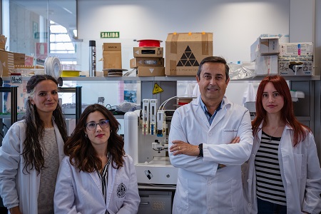
The research team belongs to the group "Advanced Therapies: differentiation, regeneration and cancer" directed by Juan Antonio Marchal, who is also part of the Biosanitary Research Institute of Granada (ibs.Granada), and of the Unit of Excellence "MNat-Modeling nature: from nano to macro” from the University of Granada. The work has been financed by the Carlos III Health Institute through a technological development project in health, by the Department of University, Research and Innovation and the Ministry of Health and Consumption, as well as by the Doctors Galera and Requena Chair of Cell Research cancer mother of the UGR. The results of this research have led to the publication in the prestigious journal Biofabrication and the registration of the patent PCT/EP2023/052738.
This research represents a breakthrough in the fight against malignant melanoma, one of the most aggressive and difficult-to-treat skin cancers, and one of the fastest growing cases diagnosed each year. In addition, thanks to the results obtained, new possibilities are opened to improve the efficacy of treatments, direct and personalize antitumor therapies and, ultimately, save lives.
Bibliographic references:
López de Andrés J, Ruiz-Toranzo M, Antich C, Chocarro-Wrona C, López-Ruíz E, Jiménez G, Marchal JA. Biofabrication of a tri-layered 3D-bioprinted CSC-based malignant melanoma model for personalized cancer treatment. Biofabrication. 2023 May 15;15(3). doi: 10.1088/1758-5090/ac8dc6.
Patent: PCT/EP2023/052738. Marchal JA, Jiménez G, López de Andrés J. “Biofabrication of a tri-layered 3D-bioprinted CSC-based malignant melanoma model”

La mejor actitud que podemos adoptar es la de trat...

The research team observed changes in head circumf...

AtCDF3 gene induced greater production of sugars a...

En nuestro post hablamos sobre este interesante tipo de célula del...

Telum Therapeutics, a biotechnology company leveraging proprietary met...


