Researchers at the Centre for Genomic Regulation (CRG) in Barcelona have identified a potential new diagnostic marker that predicts the successful and efficient development of mammalian egg cells. The findings could pave the way for generating artificial oocytes in the laboratory, helping researchers study the causes and treatments of infertility disorders and test the impact of drugs and chemicals on women’s reproduction. The research is published in The EMBO Journal.
Humans have 23 pairs of chromosomes. Between males and females, 22 pairs are shared, with the 23rd pair being the sex chromosomes. Males usually have an X and a Y chromosome, while females have two X’s. This presents a potential problem for the female cellular machinery, as two active X chromosomes generate an overdose of gene products, which is fatal for developing embryos or leads to cancer in adult life. To avoid this scenario, female cells inactivate one X chromosome by turning off its genes and compacting it.
Little is known about how X-chromosome inactivation affects the development of reproductive cells. In mammals, oocytes develop from germ cells, precursor cells that migrate from early embryonic tissue to the developing gonads. Germ cells then undergo meiosis, an important chromosomal rearrangement process, which is responsible for the genetic uniqueness of each individual germ cell. Germ cells mature and eventually turn into functional sperm or oocytes.
To address this question, researchers at the Centre for Genomic Regulation (CRG) built an X-chromosome reporter system (XRep), a tool which allowed them to study how the chromosome shapeshifts over time during germ cell development in vitro.
Using female mouse cells, the method revealed a carefully orchestrated act of X-chromosome ‘yoyo’. If one X chromosome briefly inactivated and then reactivated, it resulted in germ cells being four times more efficient for entering meiosis and transforming into egg cells compared to germ cells that have never turned their X chromosome ‘off and on’ again. In comparison, germ cells that failed to inactivate the X chromosome in the first place or reactivated it too rapidly showed abnormal gene expression and cell differentiation patterns.
The study also found that cells with two active X chromosomes divided faster and easily reverted into a state of pluripotency. According to the authors, these characteristics are similar to human germ cell tumors, which come from germ cells that got lost during their migration to the gonads or failed to differentiate properly once inside the testicles and ovaries. The researchers deduced that a correct X-chromosome inactivation and reactivation sequence is an indicator of normal germ cell differentiation. The research team notes that further studies will be needed to confirm whether an abnormal X-chromosome state is a diagnostic indicator, or whether it could be a causal factor responsible for the cell abnormality.
“Our findings have important implications for reproductive research because XRep allows us to assess cellular X-chromosome status in real time, helping identify and isolate germ cells with a high success rate of turning into oocytes,” says Dr. Bernhard Payer, Group Leader at the CRG and senior author of the study.
“Human oocytes have never been generated completely in vitro. Monitoring the X-chromosome state during in vitro germ cell differentiation might therefore be a way to optimize the protocol to produce also high quality human eggs in the lab. Human eggs for research are scarce and difficult to obtain because they are currently only available from egg donors and mostly used for reproductive purposes. In vitro-generated human eggs could therefore provide an unlimited resource to study the causes and treatments of infertility disorders and as well to test the safety of drugs and chemicals for women’s reproduction”, concludes Dr. Payer.
Dr. Moritz Bauer, co-first author of the study, adds: “Our results also highlight how we need specific tools to study female cells. The vast majority of studies are performed using male cells, leading to a gender-gap in scientific knowledge. We therefore need to stop looking at female development through the lens of male cells, which will contribute to our understanding of sex-specific disease progressions.”
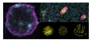
Image: an artificial ovary, where in vitro-generated female germ cells (magenta) enter meiosis (yellow cells) to become oocytes with the help from supporting cells (cyan). This process is 4-times more efficient when the X chromosome is correctly turned OFF and ON again during germ cell development. The top right panel shows oocytes at different maturation stages: from more immature on the left to more advanced on the right. The lower right panel shows the chromosomal organization during the meiosis process when germ cells become unique by reshuffling their genetic material. Credit: Dr. Jacqueline Severino/CRG

La mejor actitud que podemos adoptar es la de trat...

The research team observed changes in head circumf...

AtCDF3 gene induced greater production of sugars a...

En nuestro post hablamos sobre este interesante tipo de célula del...

Telum Therapeutics, a biotechnology company leveraging proprietary met...


