The embryonic stem cells that form faces – neural crest cells – use an unexpected mechanism to develop our facial features, according to a new UCL-led study involving IBEC researchers.
By identifying how these cells move, the researchers’ findings could help understand how facial defects, such as cleft palate and facial palsy, occur.
This newly described mechanism is likely to be found in other cell movement processes, such as cancer invasion during metastasis or wound healing, so the findings may pave the way to developing a range of new therapies for these, too.
“Neural crest cells in the womb migrate from the back to the front of the head in order to form the face,” says Prof. Roberto Mayor at University College London’s Cell & Developmental Biology department, whose group carried out the study published today in Science alongside IBEC group leader and ICREA researcher Xavier Trepat. “For the first time, we’ve identified how this migration happens: it appears to be similar to how you would squeeze toothpaste from the back of a tube to move the contents to the front.”
The traditional explanation of how cells move thought that the process was similar to a train: there’s an engine at the front that generates force and pulls the rest of the train forward. The surprising new discovery that the engine that moves neural crest cells is at the back rather than the front has important consequences, as any modification of cell movement aiming to repair facial malformation, improve wound healing or inhibit cancer metastasis should be focused on modifying the back cells rather than the front ones.
For the study, the researchers investigated embryos of both frogs and fish, because their neural crest cells behave in a similar way to those of humans and their movement is often used to study the spread of cancer. In addition, the embryo development of frogs and fish can be studied without inflicting harm.
The team used light to control the behaviour of the neural crest cell cluster using a technique called optogenetics. After identifying a protein cable surrounding the cluster that contracts to move the cluster, they found that when neural crest cells at the back of the embryo were illuminated with a laser beam, they contracted and led to movement towards the face. “This is a great example of supracellular organization, where a group of cells behaves coordinately like a single one,” says Prof. Trepat.
The study was a collaboration of researchers at UCL and IBEC, a centre of the Barcelona Institute of Science and Technology. It was funded by the Medical Research Council, the Biotechnology and Biological Sciences Research Council, the Wellcome Trust, the European Commission, ERC, MINECO, the Generalitat de Catalunya and CIBER-BBN.
—
Source article: Adam Shellard, András Szabó, Xavier Trepat & Roberto Mayor (2018). Supracellular contraction at the rear of neural crest cell groups drives collective chemotaxis. Science, Vol. 362, Issue 6412, pp. 339-343
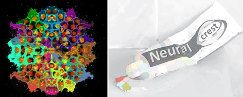
Image: Left: Neural crest migrate collectively during morphogenesis in the head; right, the cells are pushed forward from the back, like squeezing toothpaste out of a tube.

La mejor actitud que podemos adoptar es la de trat...

The research team observed changes in head circumf...

AtCDF3 gene induced greater production of sugars a...

En nuestro post hablamos sobre este interesante tipo de célula del...

Telum Therapeutics, a biotechnology company leveraging proprietary met...


